Bones Of The Foot Drawing
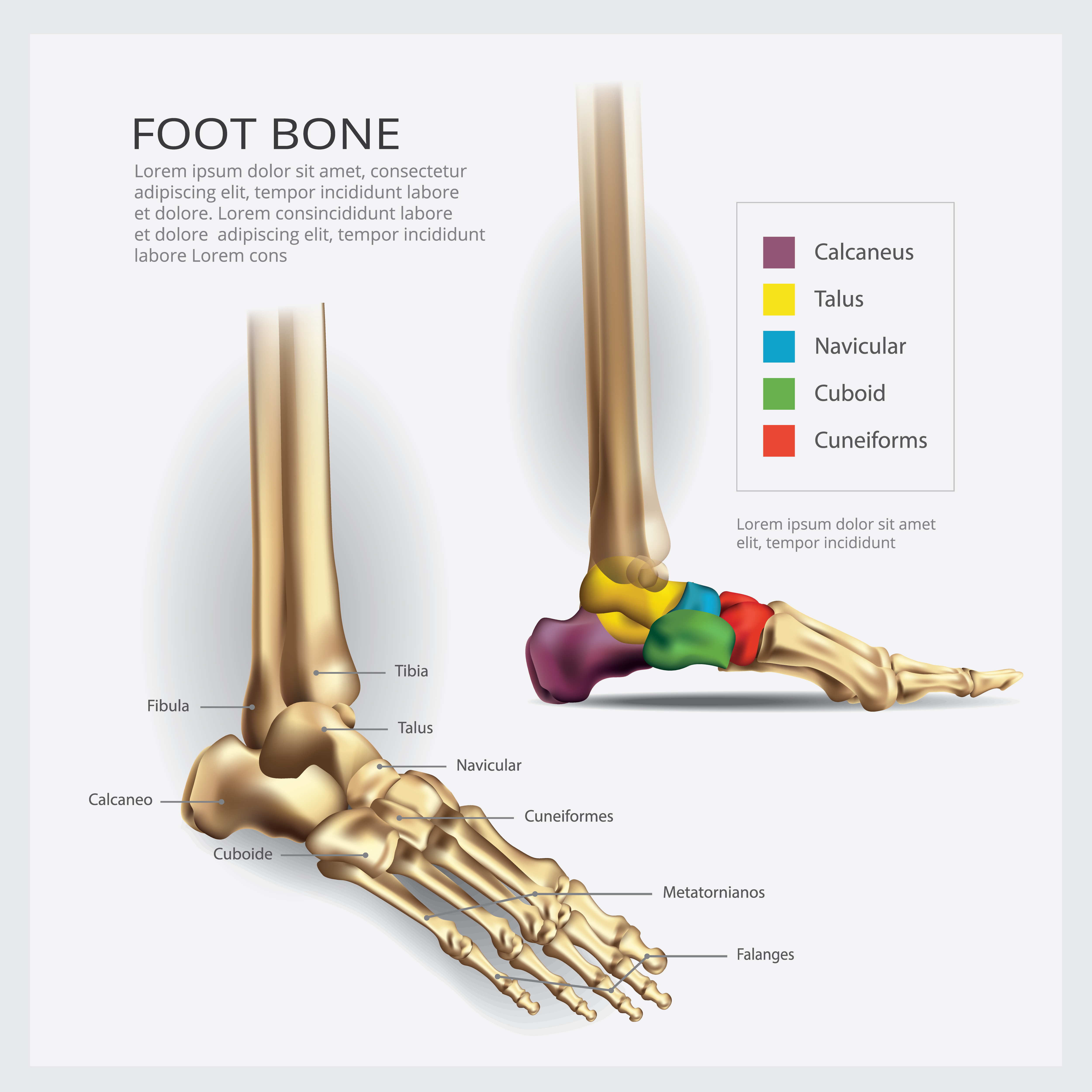
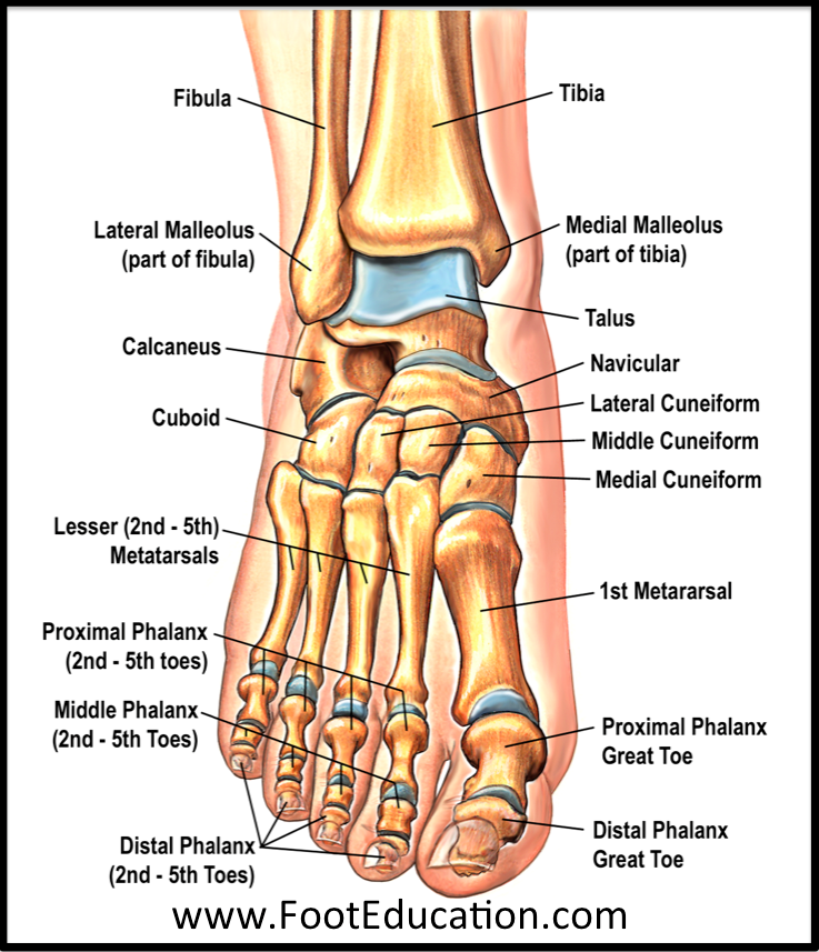
Bones Of The Foot Drawing - Web ankle joint explore study unit joints and ligaments of the foot explore study unit bones of the foot there are 26 bones in the foot, divided into three groups: Bones and landmarks covered include achilles tendon, calcaneus, phalanges, metatarsals, wrinkles, and veins. The foot can be divided into three regions, the hindfoot, midfoot, and forefoot. You'll find still images of foot bone poses in the download below. See foot anatomy drawing stock video clips filters all images photos vectors illustrations 3d objects sort by popular line drawing of the left and right foot soles. Web anatomy biology visualization muscles study leg feet foot science nerves tendons ligaments human interior bones deringungor , mert_kilicoglu , glimwork and 19 others liked this model Watch the 24 second short. Web foot bones, sketch of human anatomy. :)what's up today?today is the day for lesson.i'm going to give a lesson to draw the human foot.first, to draw the human body well, i recommend you. The midfoot is made up of five bones that form the arch of the foot. Draw from life using your own feet or draw from the 3d models i provide you. Use this as an aid in learning the names of the bones. :)what's up today?today is the day for lesson.i'm going to give a lesson to draw the human foot.first, to draw the human body well, i recommend you. The three main regions of. The hind foot consists of the heel bone, or calcaneus, and the ankle bone, or talus. Web anatomy of the foot. Web all 26 bones of the foot are described generally for drawing purposes. Web where the foot joins the leg: The foot is challenging to draw because it’s flexible, asymmetrical, and should usually look like it’s on the ground. It is level upon the ground, consistent with the heel; Drawing feet and the anatomy of them tutorial as the little finger side is the heel side of the hand, so the outside of the foot is the heel side. Web foot bones, sketch of human anatomy. You'll find still images of foot bone poses in the download below. Seven. Foot bones vector sketch of human anatomy, orthopedics medicine design. The phalanges, which are the bones in your toes. Web in this lesson, we're gonna take this simple structure that we learned about in the foot bones lesson, add the fat pads, toenails, and skin folds to draw a fleshed out foot. The hindfoot, the midfoot, and the forefoot. Web. The foot is challenging to draw because it’s flexible, asymmetrical, and should usually look like it’s on the ground (perspective). Web fortunately, the bones are a great way to study the foot. The hindfoot is the back portion of the foot, and it includes the heel, which connects the foot to the lower leg. Web the seven tarsal bones are:. The hindfoot, the midfoot, and the forefoot. Web there are 26 bones in the foot. The largest bone of the foot, it is commonly referred to as the heel of the foot. Web out of 206 bones present in the body, 28 bones are located in the human foot alone. In this article, to make it easier to undertand, the. It is level upon the ground, consistent with the heel; Use this as an aid in learning the names of the bones. Since the foot is so bony, knowing the inside. The hindfoot is the back portion of the foot, and it includes the heel, which connects the foot to the lower leg. Web the seven tarsal bones are: Foot bones vector sketch of human anatomy, orthopedics medicine design. The foot is challenging to draw because it’s flexible, asymmetrical, and should usually look like it’s on the ground (perspective). The phalanges, which are the bones in your toes. Look for it in all the diagrams above. Web all 26 bones of the foot are described generally for drawing purposes. You'll find still images of foot bone poses in the download below. In this article, to make it easier to undertand, the bones of the foot will be divided into tarsal bones, metatarsal bones, and phalanges. Drawing feet and the anatomy of them tutorial as the little finger side is the heel side of the hand, so the outside of. Web in this lesson, we're gonna take this simple structure that we learned about in the foot bones lesson, add the fat pads, toenails, and skin folds to draw a fleshed out foot. Drawing feet and the anatomy of them tutorial as the little finger side is the heel side of the hand, so the outside of the foot is. Web bone structure the bone structure of the foot consists of three different sections: The three main regions of the foot are the hindfoot, midfoot, and forefoot. Since the foot is so bony, knowing the inside anatomy directly helps you draw the outside surface. The foot is made up of 33 joints. Web anatomy biology visualization muscles study leg feet foot science nerves tendons ligaments human interior bones deringungor , mert_kilicoglu , glimwork and 19 others liked this model The foot can be divided into three regions, the hindfoot, midfoot, and forefoot. The metatarsals, which run through the flat part of your foot. Web it has three articulations: Web fortunately, the bones are a great way to study the foot. This lesson will focus on the overall design of the foot, and the form, proportion, and mobility of the individual bones. In this article, to make it easier to undertand, the bones of the foot will be divided into tarsal bones, metatarsal bones, and phalanges. :)what's up today?today is the day for lesson.i'm going to give a lesson to draw the human foot.first, to draw the human body well, i recommend you. It is the second largest bone in the. The big toe (or the hallux) has fewer bones than the other toes, and thus there is no first medial phalange. Since the foot is so bo. Web your assignment is to simplify the foot bones into their basic forms.Bones of the Feet ClipArt ETC

Foot & Ankle Bones

Structure of the human foot bone Royalty Free Vector Image
.jpg)
Foot Bone Diagram resource Imageshare

Foot Bone Anatomy Vector Illustration 539973 Vector Art at Vecteezy

Foot & Ankle Bones

Pin on Anatomy & Physiology
.jpg)
Foot Bone Diagram resource Imageshare

Bones and Joints of the Foot and Ankle Overview FootEducation

anatomy of the foot Ballet News Straight from the stage bringing
Roughly Speaking, The Front Side Of The Leg Falls Vertically Into The Foot.
Web The Seven Tarsal Bones Are:
These Bones Give Structure To The Foot And Allow For All Foot Movements Like Flexing The Toes And Ankle, Walking, And Running.
See Foot Anatomy Drawing Stock Video Clips Filters All Images Photos Vectors Illustrations 3D Objects Sort By Popular Line Drawing Of The Left And Right Foot Soles.
Related Post: