Cardiovascular System Drawing
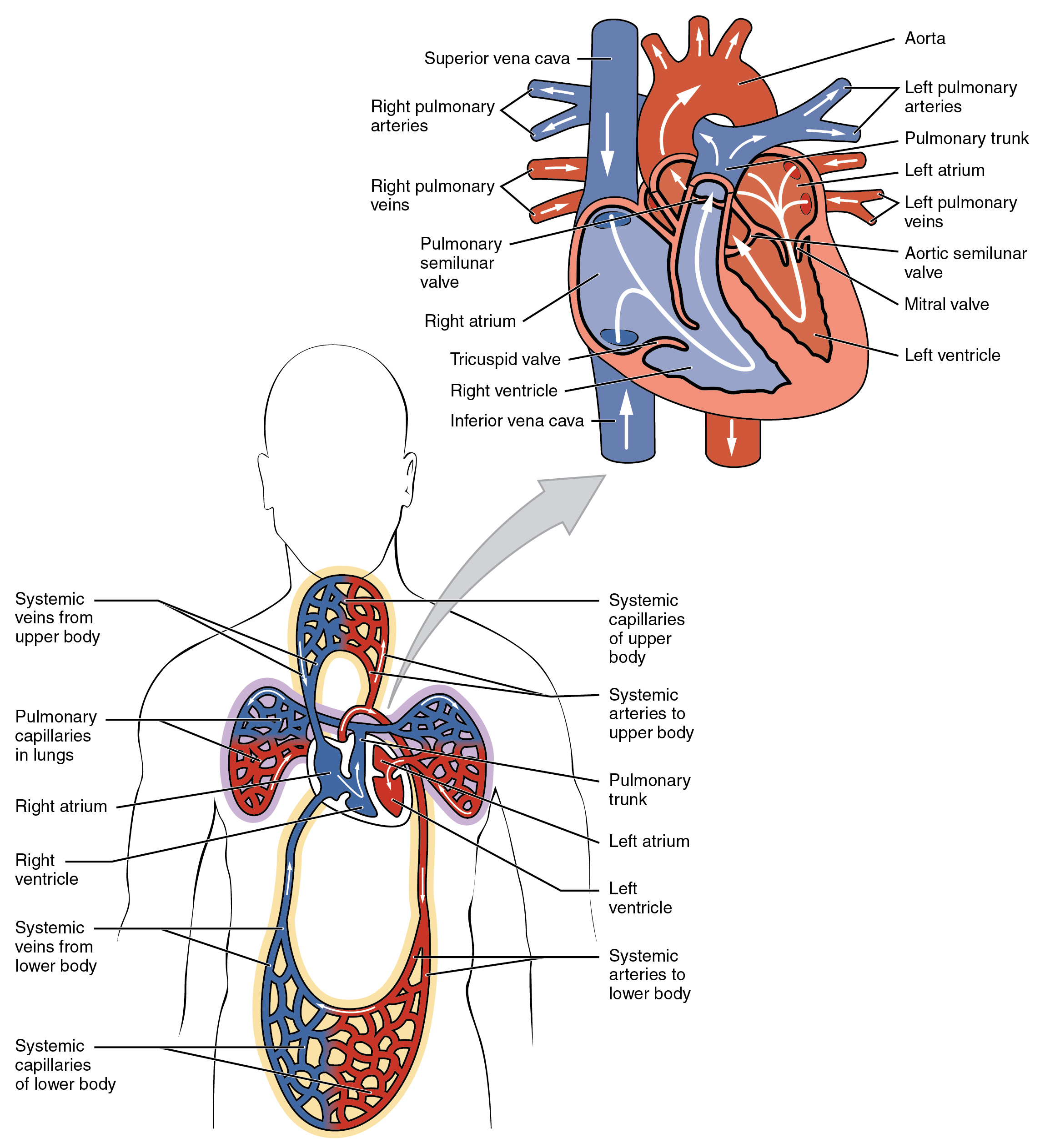
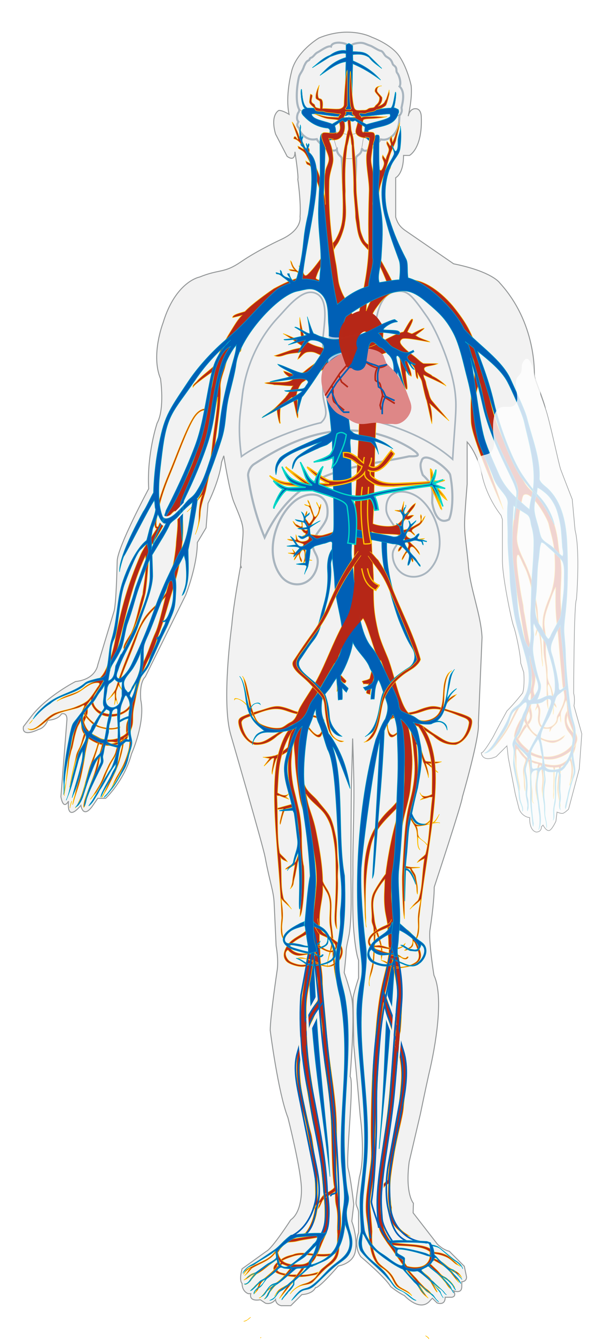
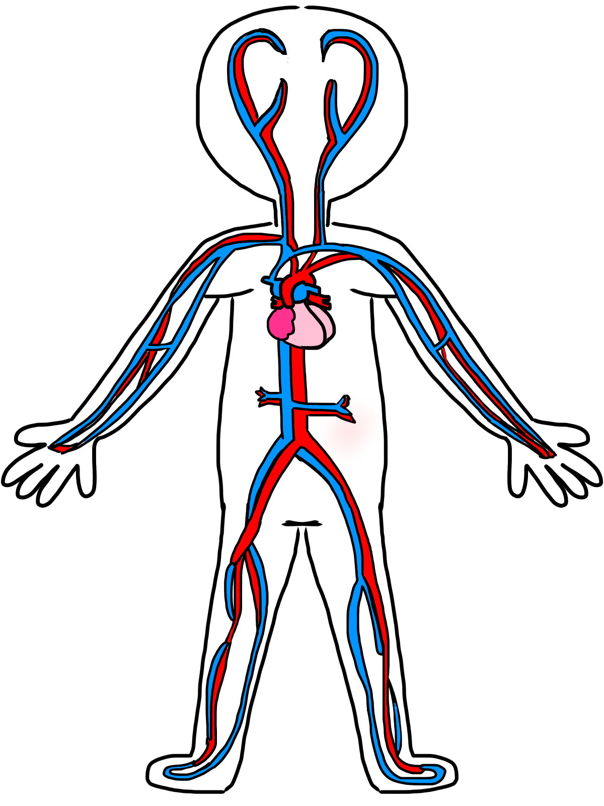
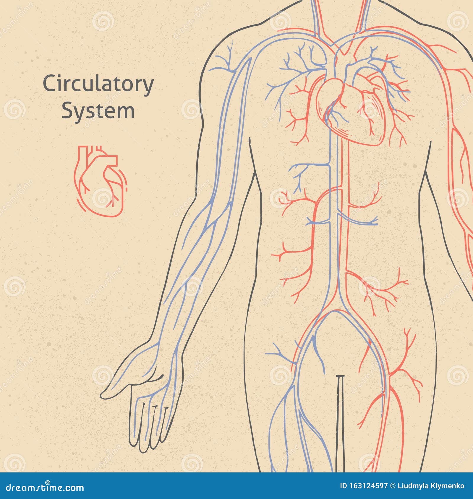
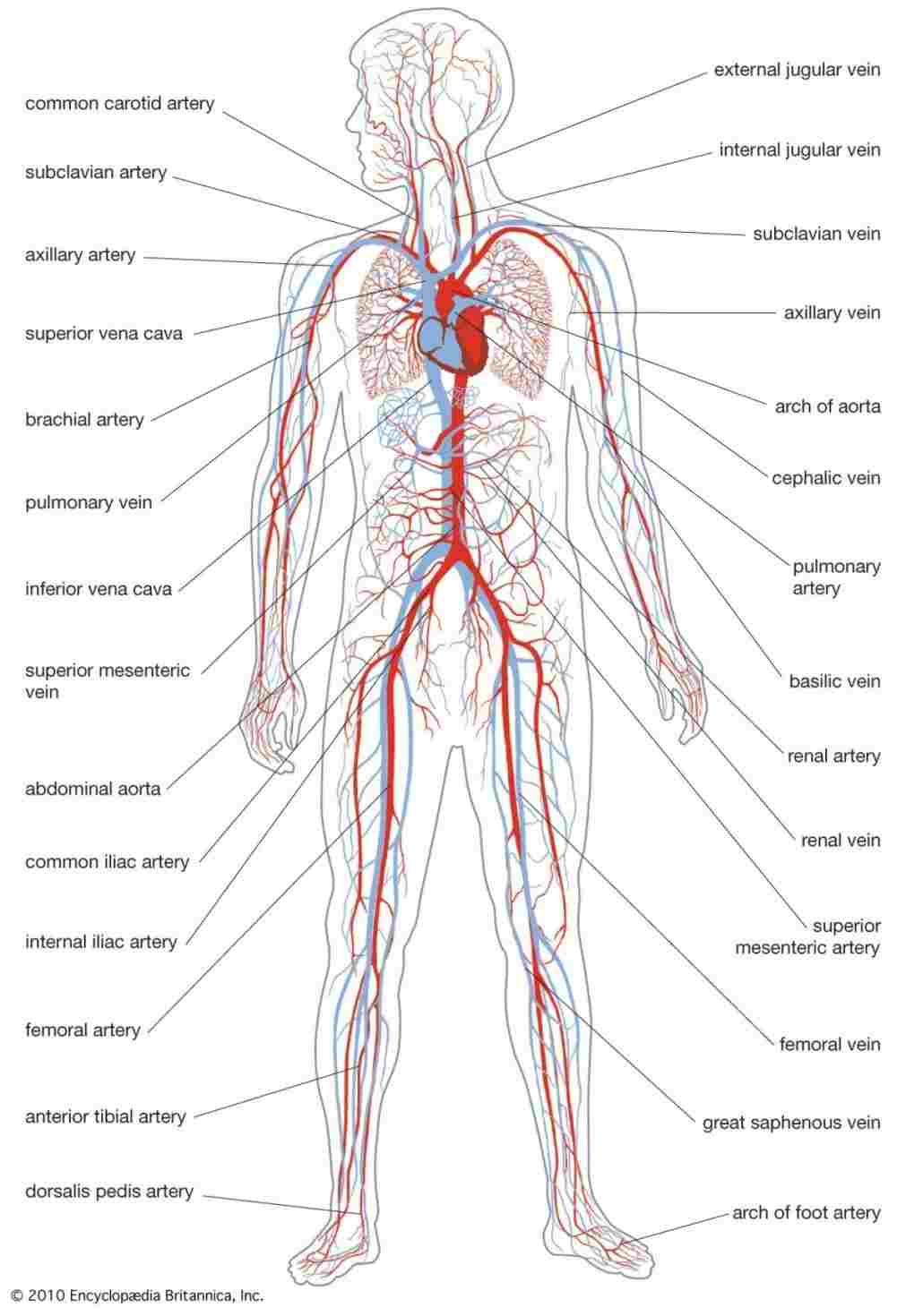
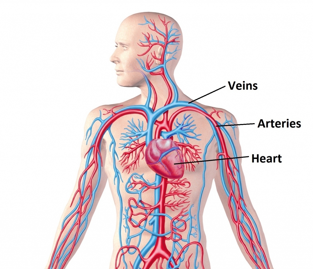
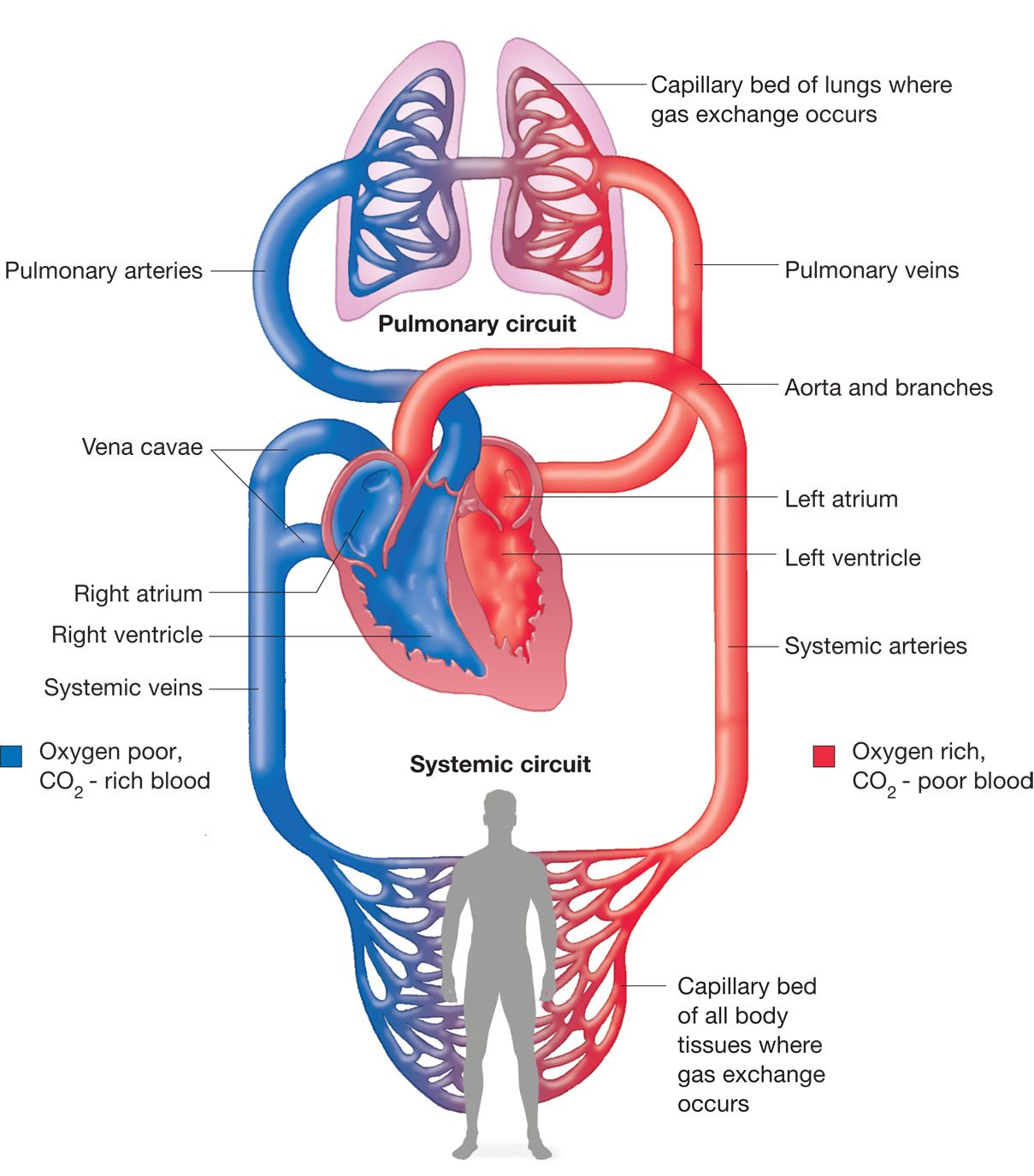
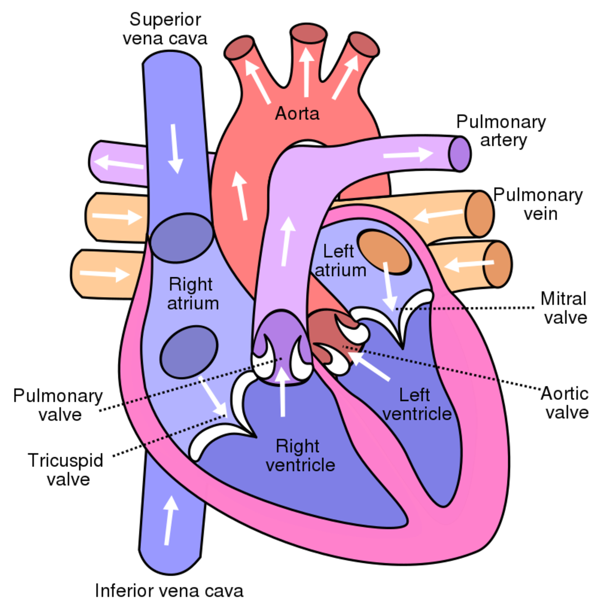
Cardiovascular System Drawing - This simplified drawing of the cardiovascular system shows its main structures. Most popular body organs poster. The human heart is located within the thoracic cavity, medially between the lungs in the space known as the mediastinum. Below is a schematic drawing showing the key structural features and relative sizes of skeletal, smooth, and cardiac muscle as you would observe them with high power. Web the cardiovascular system provides blood supply throughout the body. Figure 19.2 shows the position of the heart within the thoracic cavity. Be sure to check out our specially designed heart anatomy quiz which will help you to. The cardiovascular system consists of the heart, arteries, veins, and capillaries. Web the circulatory system, also called cardiovascular system, is a vital organ system that delivers essential substances to all cells for basic functions to occur. Doodle banners set with lungs and kidneys,. Your heart and blood vessels make up the circulatory system. Web view an interactive 3d model of the circulatory system and its major components, including the heart. Web although there are a lot of structures in the heart diagrams, you shall not worry, we’ve got them all covered for you in these articles and video tutorials. Web location of the. Web how to draw double circulatory system | step by step tutorial. It is a closed tubular system in which the blood is propelled by a muscular heart. Below is a schematic drawing showing the key structural features and relative sizes of skeletal, smooth, and cardiac muscle as you would observe them with high power. Web human cardiovascular system, organ. The right side of the heart and the left side of the heart are isolated from each other (each belongs to a separate circuit. Doodle banners set with lungs and kidneys,. The cardiovascular system consists of the heart, arteries, veins, and capillaries. Blood vessels called arteries are also shown in red, and blood vessels called veins are shown in blue.. This simplified drawing of the cardiovascular system shows its main structures. Web the heart is divided into four chambers: Web the cardiovascular system. Web the illustration depicts a circulatory system against background of a human silhouette with a place for text. Right atrium, right ventricle, left atrium, and left ventricle. #circulatorysystem #heart #lung the human circulatory system is a double circulatory system. Below is a schematic drawing showing the key structural features and relative sizes of skeletal, smooth, and cardiac muscle as you would observe them with high power. The atria are the two superior chambers of the heart and the ventricles are the two inferior chambers of the heart.. The pulmonary circuit is responsible for exchanging blood between the heart and lungs for oxygenation, while the systemic. Your heart and blood vessels make up the circulatory system. Blood vessels called arteries are also shown in red, and blood vessels called veins are shown in blue. The heart is shown in the chest in red. Web the goal of this. The heart and vessels work together intricately to provide adequate blood. Oxygenated and nutrient rich blood is distributed to tissues via the arterial system, which branches into smaller and smaller blood vessels from arteries to arterioles to capillaries (where most exchange. Web the circulatory system, also called cardiovascular system, is a vital organ system that delivers essential substances to all. Your heart and blood vessels make up the circulatory system. Web the cardiovascular system. The primary function of the heart and blood vessels is to transport oxygen, nutrients, and byproducts of metabolism. Blood vessels called arteries are also shown in red, and blood vessels called veins are shown in blue. Most popular body organs poster. Right atrium, right ventricle, left atrium, and left ventricle. Comprised of the heart, blood vessels and the blood itself, it is divided into two loops which both begin in the heart. Web location of the heart. Anatomically correct vector illustration of an internal organ in the style of engraving. Web the circulatory system consists of three independent systems that work. The heart and vessels work together intricately to provide adequate blood. Web cardiovascular system heart structure & circulation drawing to follow along with youtube text lecture notes quizlet flashcards practice questions & quizzes (with. Web although there are a lot of structures in the heart diagrams, you shall not worry, we’ve got them all covered for you in these articles. Detailed pencil drawing on an old paper. Web below is a neat labelled circulatory system diagram. Web the heart is divided into four chambers: All vertebrates have a closed circulatory system in which the blood plasma and cells remain within blood vessels. Web the goal of this lab is to examine and describe the structural organization of the heart and the major classes of blood vessels. Your heart and blood vessels make up the circulatory system. Web the circulatory system consists of three independent systems that work together: Below is a schematic drawing showing the key structural features and relative sizes of skeletal, smooth, and cardiac muscle as you would observe them with high power. Another part of the circulatory system is to remove waste from cells and organs so your body can dispose. Oxygenated and nutrient rich blood is distributed to tissues via the arterial system, which branches into smaller and smaller blood vessels from arteries to arterioles to capillaries (where most exchange. #circulatorysystem #heart #lung the human circulatory system is a double circulatory system. Right atrium, right ventricle, left atrium, and left ventricle. Web the illustration depicts a circulatory system against background of a human silhouette with a place for text. By responding to various stimuli, it can control the velocity and amount of blood carried through the vessels. Web using the pictures you took as guide, create a drawing to show the anatomical features of the heart in the space provided below on your drawing, label the right and left ventricles, the right and left atria, the bicuspid and tricuspid valves, the papillary muscles, the chordae tendineae, the interventricular septum, the pulmonary trunk and the. Anatomically correct vector illustration of an internal organ in the style of engraving.
Cardiovascular system, artwork Stock Image F006/2990 Science
Cardiovascular System Medical Terminology for Healthcare Professions
14.2 Introduction to the Cardiovascular System Human Biology
Cardiovascular System Drawing at GetDrawings Free download
Vector Illustration of the Human Circulatory System Drawn in Retro
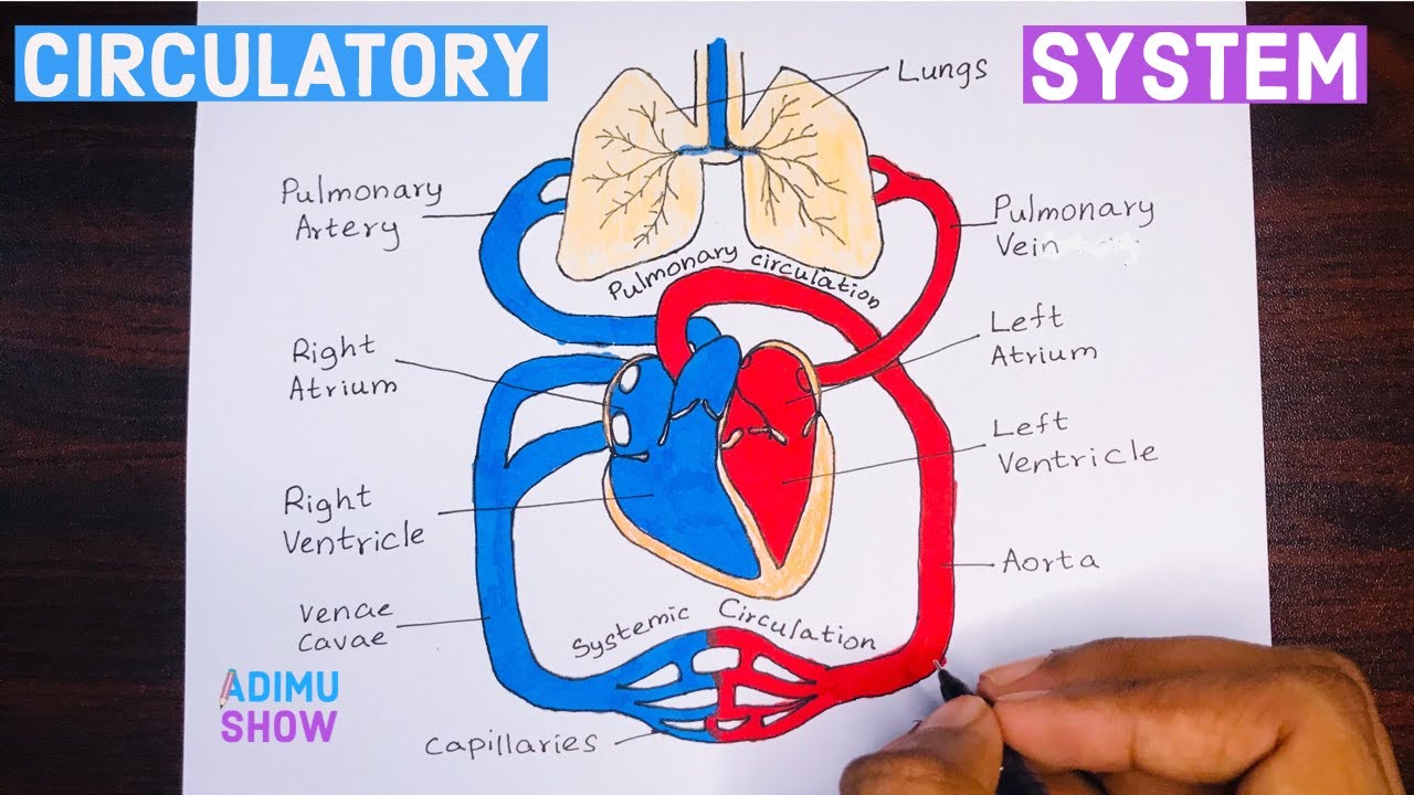
How To Draw Double circulatory System step by step tutorial YouTube
Cardiovascular System Drawing at Explore
Cardiovascular System Drawing at GetDrawings Free download
What is the Function of Veins in the Circulatory System? First Aid
The Circulatory System Its 4 Main Parts and How They Work Owlcation
The Main Function Of The Circulatory System Is To Provide Oxygen, Nutrients And Hormones To Muscles, Tissues And Organs Throughout Your Body.
The Right Side Of The Heart And The Left Side Of The Heart Are Isolated From Each Other (Each Belongs To A Separate Circuit.
The Heart And Vessels Work Together Intricately To Provide Adequate Blood.
Figure 19.2 Shows The Position Of The Heart Within The Thoracic Cavity.
Related Post: