Draw And Label A Cell
Draw And Label A Cell - Web a cell is the smallest living thing in the human organism, and all living structures in the human body are made of cells. There are two basic types of cells in nature, viz., prokaryotic cells and eukaryotic cells. To check if you have understood the cell parts, draw a blank animal cell diagram and try to fill in the different parts without referring to the labeled one given here. This will help you understand how much you have comprehended. Web draw a cell from the azolla in the space below. Highlight the inner portion of the cell known as cytoplasm with a yellow marker. Web prokaryotic cells 2.2.1 draw and label a diagram of the ultrastructure of escherichia coli (e. Web the plasma membrane not only defines the borders of the cell, but also allows the cell to interact with its environment in a controlled way. They are different from plant cells in that they do contain cell walls and chloroplast. Coli) as an example of a prokaryote. The cell contains many cell parts with different shapes. Most cells are covered by a protective membrane known as the cell wall which gives the cells their shape and rigidity. They're also the more complex of the two. Learn about the similarities and differences between plant and animal cells as we compare and contrast. Cells must be able to exclude,. Web draw a cell from the azolla in the space below. This will help you understand how much you have comprehended. Draw this animal cell by following this drawing lesson. Web label the cell and describe the process by which proteins are made and then exported. Using a cell diagram as the reference point, these quizzes challenge you to label. There are hundreds of different types of cells in the human body, which vary in shape (e.g. Web label the cell and describe the process by which proteins are made and then exported. A single cell is often a complete organism in itself, such as a bacterium or yeast. Other cells acquire specialized functions as they mature. Label the cell. They're also the more complex of the two. Web a cell is the smallest living thing in the human organism, and all living structures in the human body are made of cells. There are two basic types of cells in nature, viz., prokaryotic cells and eukaryotic cells. Web label the cell and describe the process by which proteins are made. Web prokaryotic cells 2.2.1 draw and label a diagram of the ultrastructure of escherichia coli (e. Also, draw an outer circle with a darker shade. There are hundreds of different types of cells in the human body, which vary in shape (e.g. Web the plasma membrane not only defines the borders of the cell, but also allows the cell to. Web a comparison of plant and animal cells using labelled diagrams and descriptive explanations. Highlight the inner portion of the cell known as cytoplasm with a yellow marker. Label the parts of the plant and animal cell is shared under a not declared license and was authored, remixed, and/or curated by libretexts. Protects the cell from the outside environment and. The layers in an onion bulb are fleshy leaves that have been modified to store starches for the plant. Begin by making a big circle and lines connecting it to label the cells. Comparison of prokaryotic cells and eukaryotic cells: Web in animal cells, cytokinesis is contractile, pinching the cell in two like a coin purse with a drawstring. Web. There are a number of rules/conventions that are followed when making a biological drawing. Cells must be able to exclude, take in, and excrete various substances, all in specific amounts. The layers in an onion bulb are fleshy leaves that have been modified to store starches for the plant. Also, draw an outer circle with a darker shade. The “drawstring”. The cell structure illustrations for these diagrams were generated in biorender. You can make the circle misshapen or oblong. Highlight the inner portion of the cell known as cytoplasm with a yellow marker. Most cells are covered by a protective membrane known as the cell wall which gives the cells their shape and rigidity. Web how to draw a animal. Protects the cell from the outside environment and maintains the shape of the cell.it also prevents the cell from bursting if internal. The animal cell diagram is widely asked in class 10 and 12 examinations and is beneficial to understand the structure and functions of an animal. Using a cell diagram as the reference point, these quizzes challenge you to. Most cells are covered by a protective membrane known as the cell wall which gives the cells their shape and rigidity. Web draw a cell from the azolla in the space below. Draw a simple circle or oval for the cell membrane. Prokaryotes are the simplest cells without a nucleus and cell organelles. Web how to draw a animal cell easy and step by step. Coli) as an example of a prokaryote. Web a comparison of plant and animal cells using labelled diagrams and descriptive explanations. Web the outermost part of the cell, which is shown as an outline of the cell, is labeled cell membrane. They are different from plant cells in that they do contain cell walls and chloroplast. Round, flat, long and thin, short and thick) and size (e.g. You can make the circle misshapen or oblong. The cell membrane of an animal cell is not a perfect circle. Web draw a starfish egg with a diameter of approximately 2 cm. The important part is that it does not have any sharp edges. The animal cell diagram is widely asked in class 10 and 12 examinations and is beneficial to understand the structure and functions of an animal. Begin by making a big circle and lines connecting it to label the cells.
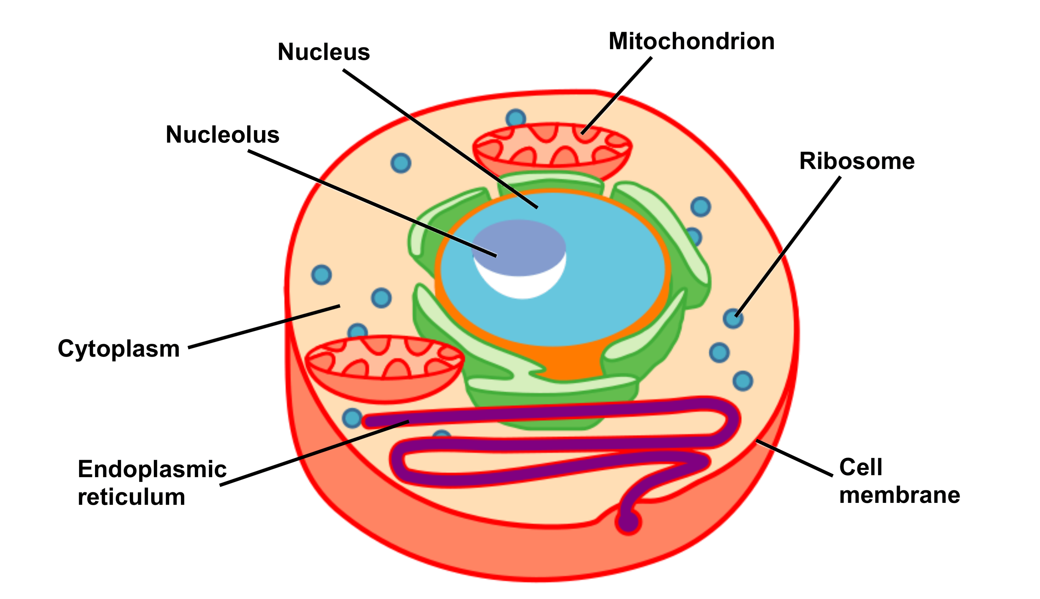
Explain the nucleus of a cell with a neat labeled diagram Science

Lysosome In Animal Cell

How to Draw a Plant Cell Biology YouTube

plantcelldiagram Tim's Printables

What is a cell? Facts
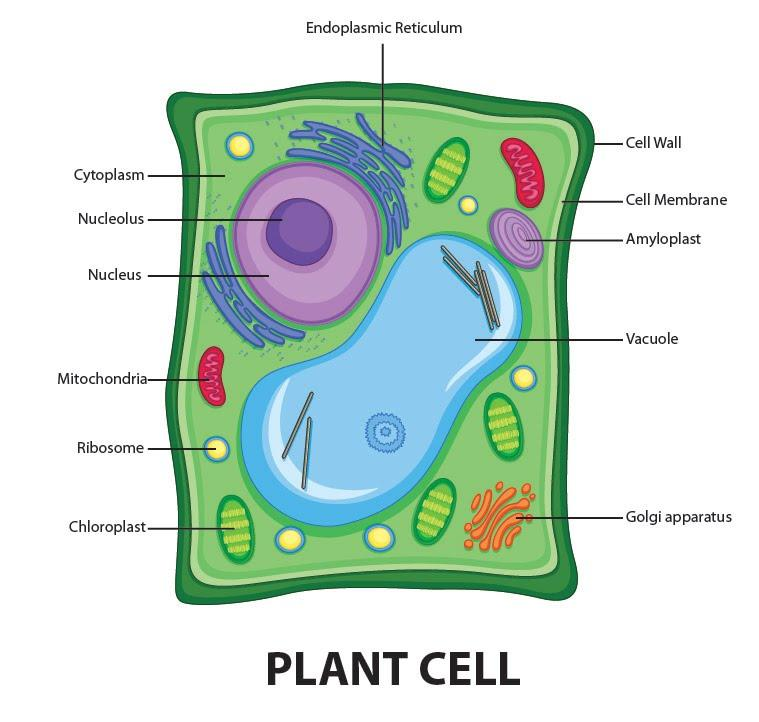
Draw a welllabelled diagram of a plant cell.

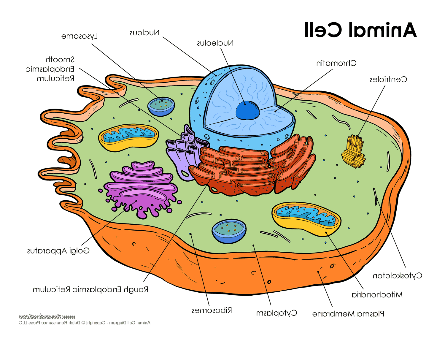
Animal Cell Diagram drawing How To Draw Animal Cell Labeled Science
Cell Structure and Function Part 1 The Organelles Medical Exam Prep
Draw A Labelled Diagram Of Plant Cell And Animal Cell Books and News

Animal Cell Parts Easy Drawing / Draw It Neat How To Draw Animal Cell
There Are A Number Of Rules/Conventions That Are Followed When Making A Biological Drawing.
A Single Cell Is Often A Complete Organism In Itself, Such As A Bacterium Or Yeast.
Web The Plasma Membrane Not Only Defines The Borders Of The Cell, But Also Allows The Cell To Interact With Its Environment In A Controlled Way.
The Cell Structure Illustrations For These Diagrams Were Generated In Biorender.
Related Post: