Drawing Of The Pancreas
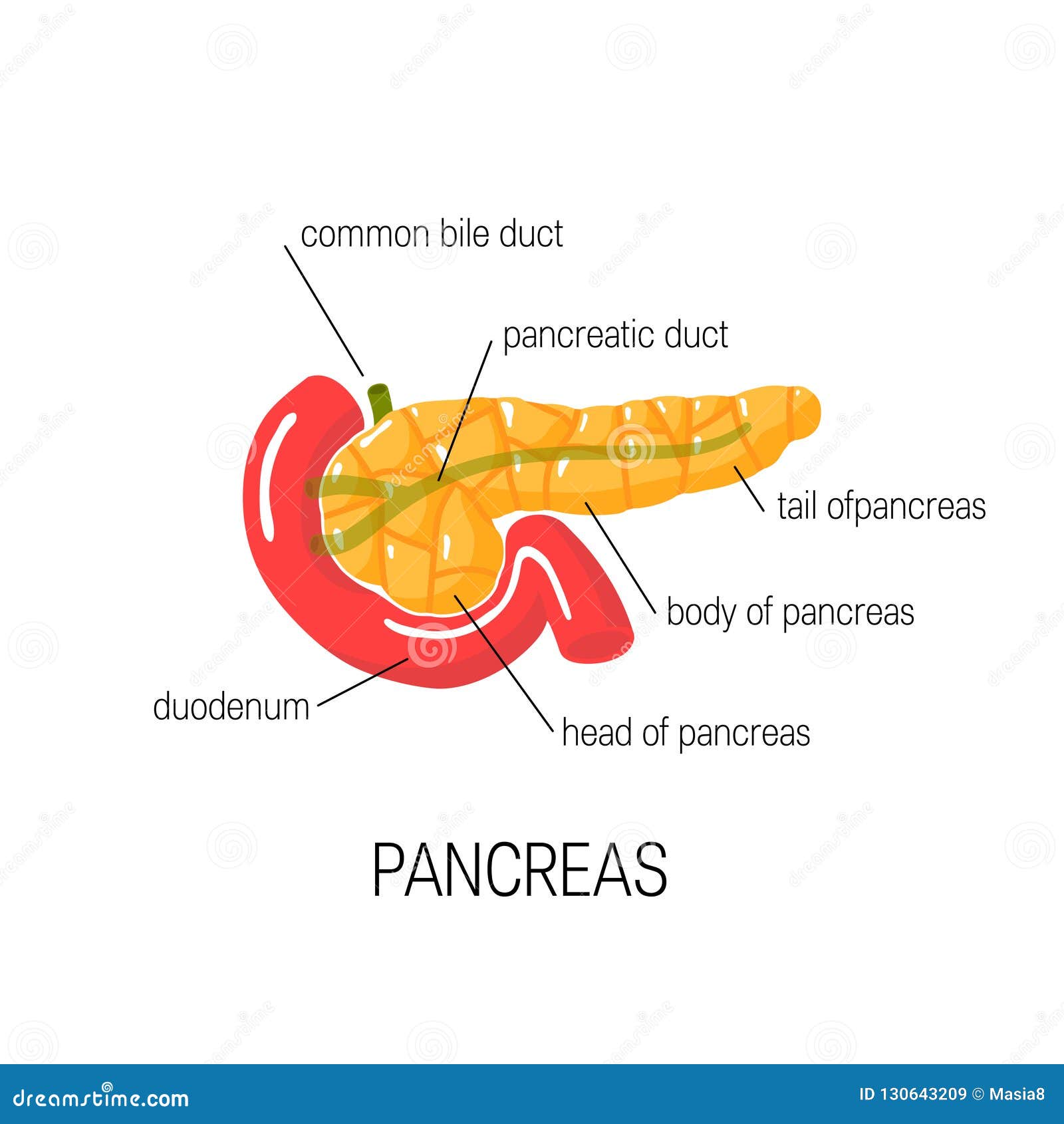
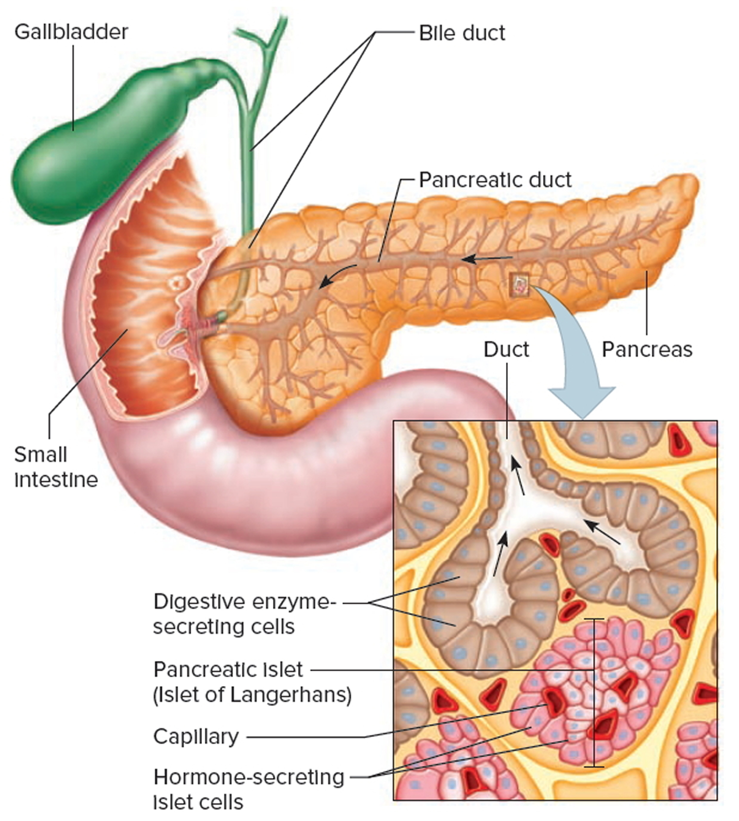
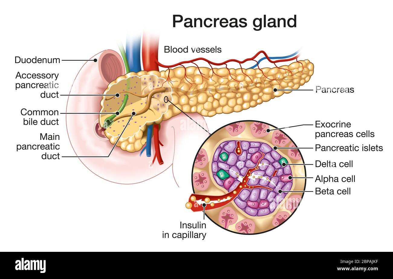
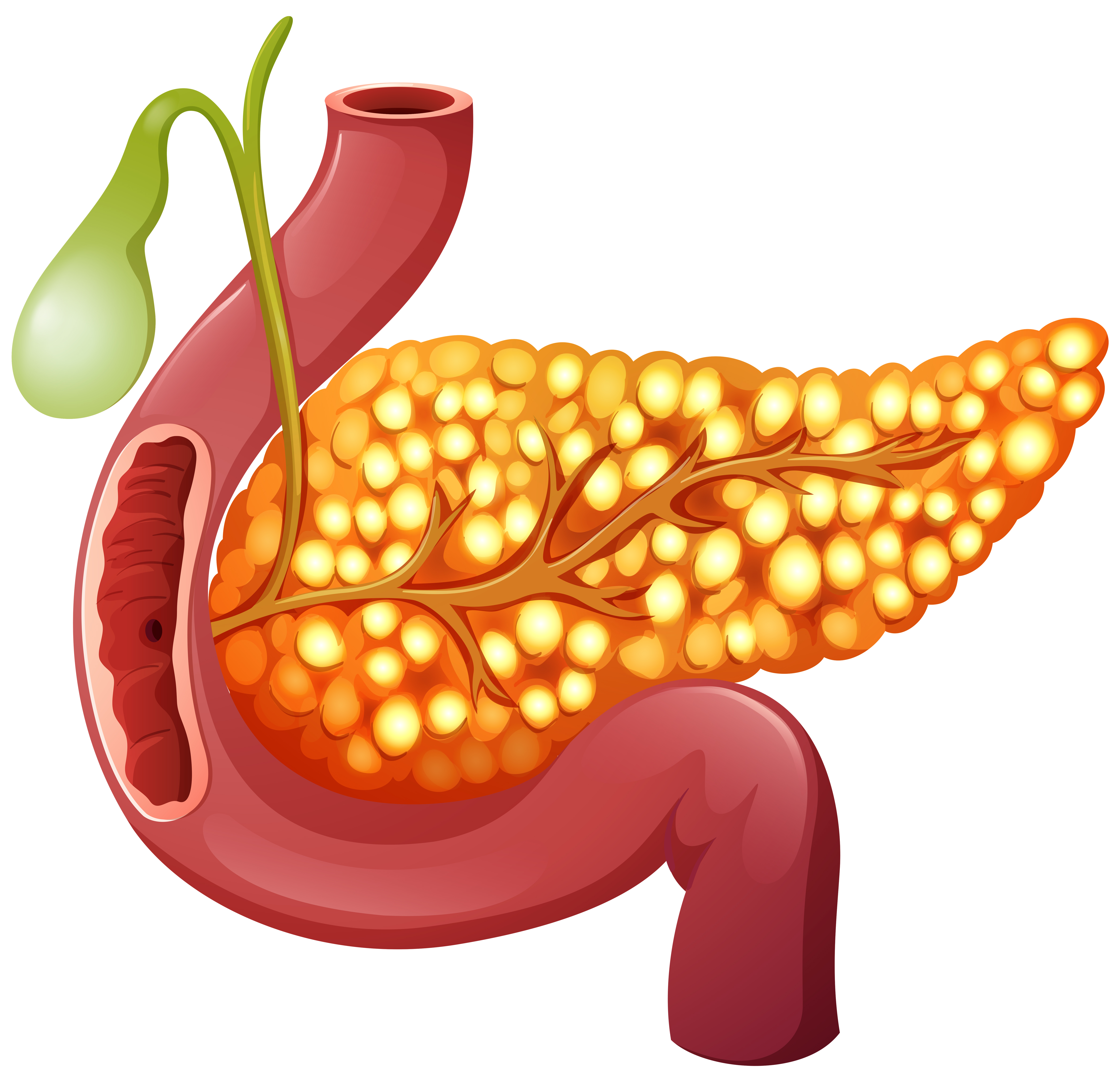
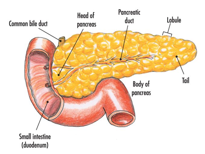
Drawing Of The Pancreas - Web the images range from classic work of skilled medical artists to original drawings and photomicrographs from leaders in the study of pancreatic anatomy and structure. Produces substances (enzymes) that help with digestion. It sits within the curve of the duodenum (the first part of the small intestine) and is divided into two parts: Web what is the pancreas? This will teach you how to draw pancreas diagram easily. Web the pancreas is an elongated gland located deep within the abdomen, tucked in between the stomach and the spine. One end of the pancreas is wider than the other and is called the head: Web pancreas is a long, slender organ, most of which is located posterior to the bottom half of the stomach ( figure 17.9.1 ). These stained samples can then be examined for drawing and labelling to identify the exocrine and endocrine tissues of the pancreas Bodytomy elaborates more on the anatomy of the human pancreas. It forms an integral part of the digestive system. The pancreas is located below and behind the stomach. One end of the pancreas is wider than the other and is called the head: It is part of your digestive system. Schematic drawings of the successive stages in the development of the pancreas from the fifth through the eighth weeks. It can be divided into 4 parts―the head, neck, body, and the tail. It sits within the curve of the duodenum (the first part of the small intestine) and is divided into two parts: Glands are organs that produce and release substances in the body. The location of the pancreas is mostly retroperitoneal, except for the tail. Web the images. One end of the pancreas is wider than the other and is called the head: To put it in a clinical context, its oblique position makes it impossible to see the entire pancreas in a single transverse section. It can be divided into 4 parts―the head, neck, body, and the tail. Exocrine glandular tissues in the pancreas produce pancreatic enzymes. Anterior to the pancreas are the stomach, colon, omentum (more.) #pancreas #howtodraw #adimushowthis is an easy drawing of pancreas. Liver insulin diabetes pancreatic cancer varicose veins pancreas icon pancreas illustration pancreas cancer 3d pancreas pancreas anatomy diabetes pancreas pancreas 3d This will teach you how to draw pancreas diagram easily. Web the pancreas is a large, mixed gland composed of. Additional useful images are available online at other websites. Growth and rotations (arrows) of the duodenum bring the ventral pancreatic bud toward the dorsal. Web choose from drawing of pancreas stock illustrations from istock. Web the pancreas is a glandular organ that produces a number of hormones essential to the body. One end of the pancreas is wider than the. Web the pancreas is an elongated organ (approximately 15 cm) which lies obliquely across the posterior abdominal wall, at the level of the l1 and l2 vertebral bodies. It forms an integral part of the digestive system. The endocrine portion is arranged as discrete islets of langerhans, which are composed of five different endocrine cell types (alpha, beta, delta, epsilon,. It serves both exocrine and endocrine functions. The head, uncinate process, neck, body and tail. Drawing shows the pancreas, stomach, spleen, liver, bile ducts, gallbladder, small intestine, and colon. Cute funny pancreas waving hand character. Only show results related to: Although it is primarily an exocrine gland, secreting a variety of digestive enzymes, the pancreas also has endocrine cells. The location of the pancreas is mostly retroperitoneal, except for the tail. An inset shows the head, body, and tail of the pancreas and the bile duct and pancreatic duct. This will teach you how to draw pancreas diagram easily. One. The pancreas is an organ and a gland. Drawing shows the pancreas, stomach, spleen, liver, bile ducts, gallbladder, small intestine, and colon. It forms an integral part of the digestive system. This cartoon represents the anatomical features of a “slice” of the abdomen at the level depicted in the upper right hand corner of the figure. Web choose from drawing. One end of the pancreas is wider than the other and is called the head: These stained samples can then be examined for drawing and labelling to identify the exocrine and endocrine tissues of the pancreas Cute funny pancreas waving hand character. Web the pancreas is an elongated organ (approximately 15 cm) which lies obliquely across the posterior abdominal wall,. Cute funny pancreas waving hand character. Vector hand drawn traditional cartoon vintage, retro, kawaii character illustration icon. Web pancreas is a long, slender organ, most of which is located posterior to the bottom half of the stomach ( figure 17.9.1 ). The pancreas is an organ in the back of your abdomen (belly). Web the pancreas is a composite organ, which has exocrine and endocrine functions. An inset shows the head, body, and tail of the pancreas and the bile duct and pancreatic duct. The pancreas performs two main functions: Web the histology of the pancreas can be studied by staining sections of pancreatic tissue and viewing them under a microscope; Drawing shows the pancreas, stomach, spleen, liver, bile ducts, gallbladder, small intestine, and colon. The head, uncinate process, neck, body and tail. The head proper and the uncinate process. Additional useful images are available online at other websites. To put it in a clinical context, its oblique position makes it impossible to see the entire pancreas in a single transverse section. These stained samples can then be examined for drawing and labelling to identify the exocrine and endocrine tissues of the pancreas This cartoon represents the anatomical features of a “slice” of the abdomen at the level depicted in the upper right hand corner of the figure. Liver insulin diabetes pancreatic cancer varicose veins pancreas icon pancreas illustration pancreas cancer 3d pancreas pancreas anatomy diabetes pancreas pancreas 3d
Pancreas Medical anatomy, Human anatomy and physiology, Anatomy
Medical Diagram of Pancreas, Vector Illustration Stock Vector
Pancreas Location, Anatomy and Function in Digestion
/pancreas_lg-595e8eae3df78c4eb64f2fce.jpg)
Pancreas Anatomy and Function
Medically illustration showing pancreas gland and pancreatic islets
Pancreas Anatomy Labeled Stock Illustration Download Image Now
A healthy human Pancreas 303570 Vector Art at Vecteezy

Human pancreas, illustration Stock Image F013/3615 Science Photo
Structure and functions of pancreas Online Science Notes

Illustration of a woman's pancreas Stock Image F023/5801 Science
See Pancreas Simple Drawings Stock Video Clips.
Only Show Results Related To:
Anterior To The Pancreas Are The Stomach, Colon, Omentum (More.)
It Can Be Divided Into 4 Parts―The Head, Neck, Body, And The Tail.
Related Post:
