Epithelial Tissue Drawing
Epithelial Tissue Drawing - The alveoli of lungs where gases diffuse, segments of. Simple squamous epithelium, because of the thinness of the cell, is present where rapid passage of chemical compounds is observed. Animal tissues in easy steps and compact way. If you need more epithelial tissue drawing or real slide pictures, then let me know. Web this video explains how to draw different types of epithelial tissue; The cells in this tissue are tightly packed within a thin ecm. Web you will also find the transitional epithelial tissue in the ureter. Web the endothelium is the epithelial tissue that lines vessels of the lymphatic and cardiovascular system, and it is made up of a single layer of squamous cells. The shape of the cells in the single cell layer of simple epithelium reflects the functioning of those cells. Web epithelial tissue is often classified according to numbers of layers of cells present, and by the shape of the cells. Simple squamous epithelium, because of the thinness of the cell, is present where rapid passage of chemical compounds is observed. Web you will also find the transitional epithelial tissue in the ureter. You may try to draw the same, but please follow the instruction from the books. Web this video explains how to draw different types of epithelial tissue; All. Animal tissues in easy steps and compact way. If it is a stratified epithelium draw all the layers. Web epithelial tissue is often classified according to numbers of layers of cells present, and by the shape of the cells. You may try to draw the same, but please follow the instruction from the books. Web the endothelium is the epithelial. All substances that enter the body must cross an epithelium. Forming sheets that cover the internal and external body surfaces (surface epithelium) and secreting organs (glandular epithelium). The cells in this tissue are tightly packed within a thin ecm. Web this video explains how to draw different types of epithelial tissue; The alveoli of lungs where gases diffuse, segments of. Simple squamous epithelium, because of the thinness of the cell, is present where rapid passage of chemical compounds is observed. The shape of the cells in the single cell layer of simple epithelium reflects the functioning of those cells. The cells of an epithelium act as gatekeepers of the body, controlling permeability by allowing selective transfer of materials across its. Simple squamous epithelium, because of the thinness of the cell, is present where rapid passage of chemical compounds is observed. Animal tissues in easy steps and compact way. Now, i will share some epithelial tissue drawing pictures with you. Epithelial tissues are found on the surfaces of all organs inside and out of the human body and as such, epithelial. The main functions of epithelia are protection from the environment, coverage, secretion and excretion, absorption, and filtration. Epithelial tissues provide the body’s first line of protection from physical, chemical, and biological damage. Web epithelial tissue is often classified according to numbers of layers of cells present, and by the shape of the cells. Forming sheets that cover the internal and. Web you will also find the transitional epithelial tissue in the ureter. The cells of an epithelium act as gatekeepers of the body, controlling permeability by allowing selective transfer of materials across its surface. Animal tissues in easy steps and compact way. If it is a stratified epithelium draw all the layers. Simple epithelial tissue is organised as a single. Simple squamous epithelium, because of the thinness of the cell, is present where rapid passage of chemical compounds is observed. Web the endothelium is the epithelial tissue that lines vessels of the lymphatic and cardiovascular system, and it is made up of a single layer of squamous cells. Web epithelial tissue is often classified according to numbers of layers of. Web the endothelium is the epithelial tissue that lines vessels of the lymphatic and cardiovascular system, and it is made up of a single layer of squamous cells. Now, i will share some epithelial tissue drawing pictures with you. Forming sheets that cover the internal and external body surfaces (surface epithelium) and secreting organs (glandular epithelium). Web you will also. Simple squamous epithelium, because of the thinness of the cell, is present where rapid passage of chemical compounds is observed. The alveoli of lungs where gases diffuse, segments of. You may try to draw the same, but please follow the instruction from the books. Web this video explains how to draw different types of epithelial tissue; Now, i will share. Web epithelium is one of only 4 types of human body tissues.like all types, it is formed by cells within an extracellular matrix (ecm). This video helps you to draw science diag. Web epithelial tissue is often classified according to numbers of layers of cells present, and by the shape of the cells. Epithelial tissues are found on the surfaces of all organs inside and out of the human body and as such, epithelial tissues have one edge not connected to other cells, the apical surface.the other end of an epithelial tissue (the basal surface) is attached to a basement membrane that is partly produced by the epithelial cells. Simple squamous epithelium, because of the thinness of the cell, is present where rapid passage of chemical compounds is observed. The main functions of epithelia are protection from the environment, coverage, secretion and excretion, absorption, and filtration. The cells in this tissue are tightly packed within a thin ecm. Now, i will share some epithelial tissue drawing pictures with you. Web you will also find the transitional epithelial tissue in the ureter. The alveoli of lungs where gases diffuse, segments of. Web this video explains how to draw different types of epithelial tissue; Epithelial tissues provide the body’s first line of protection from physical, chemical, and biological damage. If you need more epithelial tissue drawing or real slide pictures, then let me know. The alveoli of lungs where gases diffuse, segments of. Simple squamous epithelium, because of the thinness of the cell, is present where rapid passage of chemical compounds is observed. Forming sheets that cover the internal and external body surfaces (surface epithelium) and secreting organs (glandular epithelium).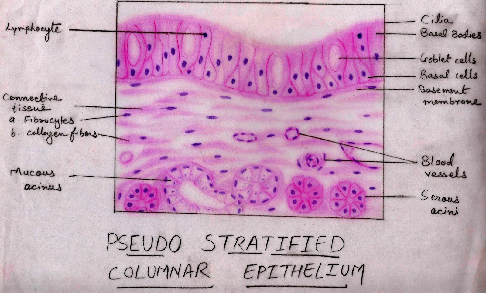
Histology Image Membranous epithelium
Epithelial tissues

epithelial tissue, drawing Stock Image C015/2525 Science Photo
Tissues Basicmedical Key
Epithelial Tissue Characteristics, Types, and Functions Owlcation
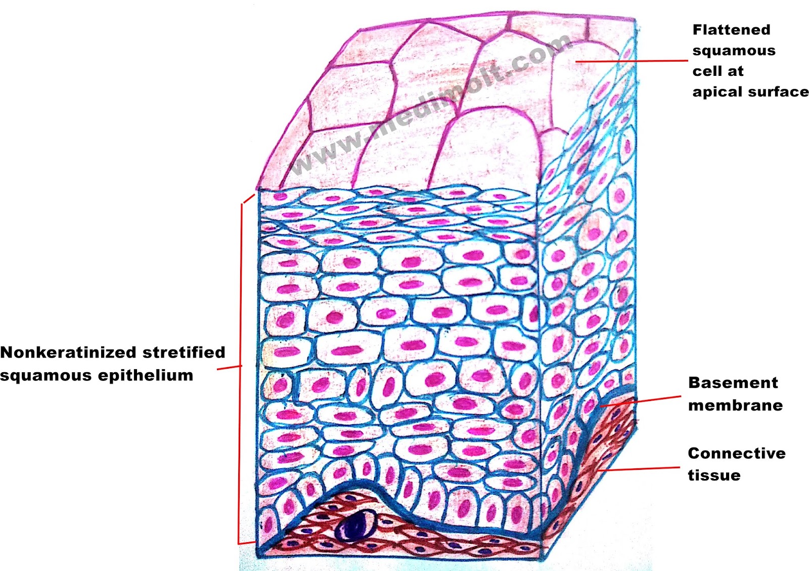
Simple Squamous Epithelium Basement Membrane Esophagus Stratified Squamous
Epithelial Cells
Pseudostratified Columnar Epithelium Diagram
Describe various types of epithelial tissues with the help of labeled
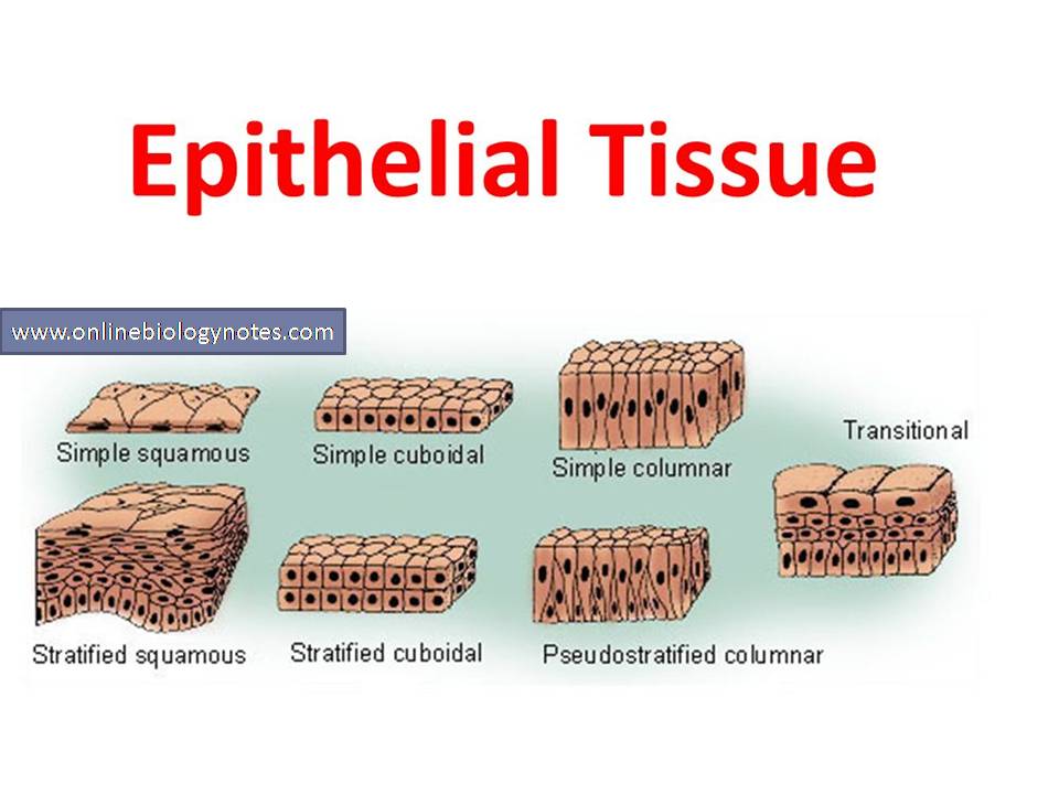
Epithelial tissue characteristics and classification scheme and types
Web In Epithelial Tissue, Cells Are Closely Packed With Little Or No Extracellular Matrix Except For The Basal Lamina That Separates The Epithelium From Underlying Tissue.
All Substances That Enter The Body Must Cross An Epithelium.
If It Is A Stratified Epithelium Draw All The Layers.
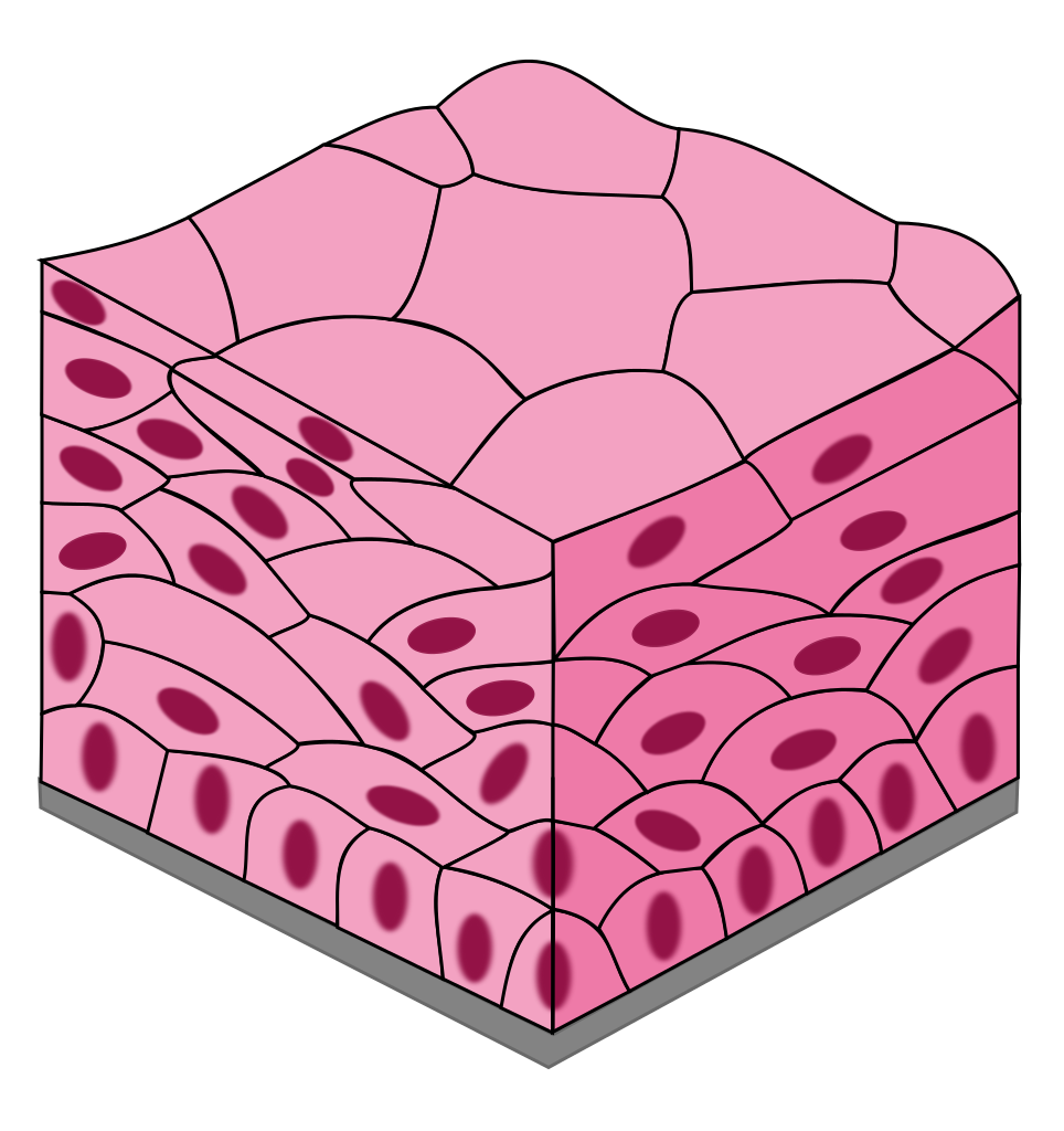
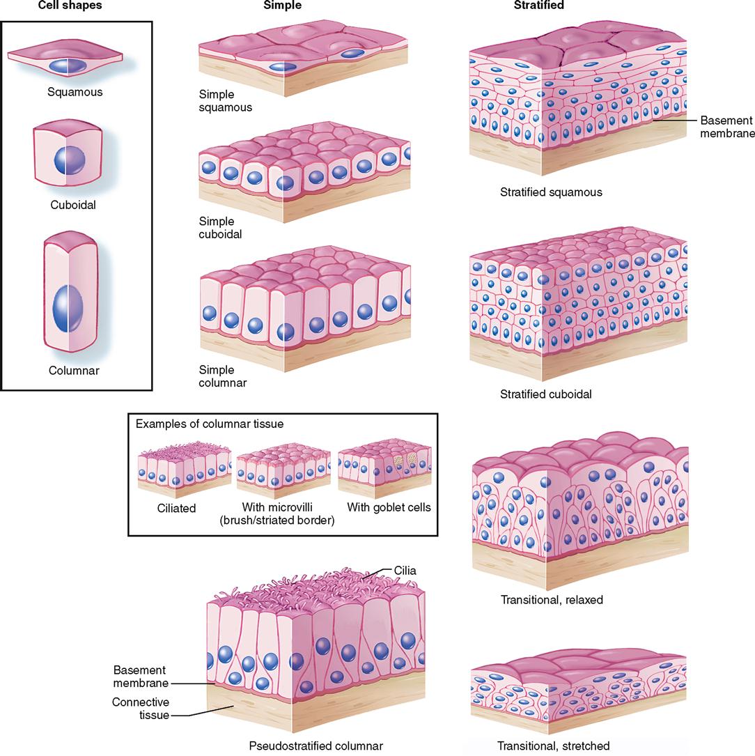
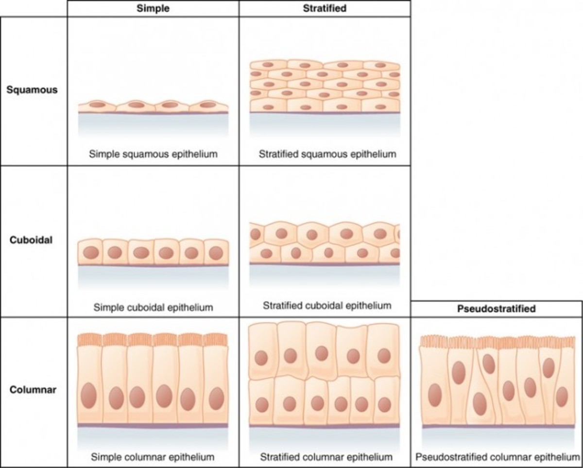
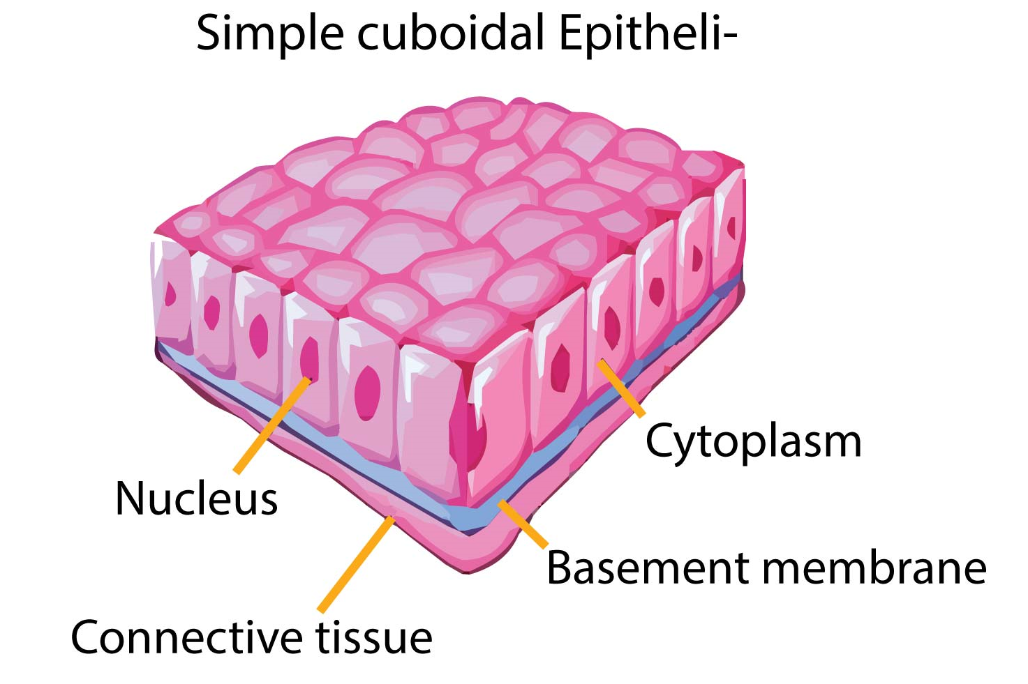
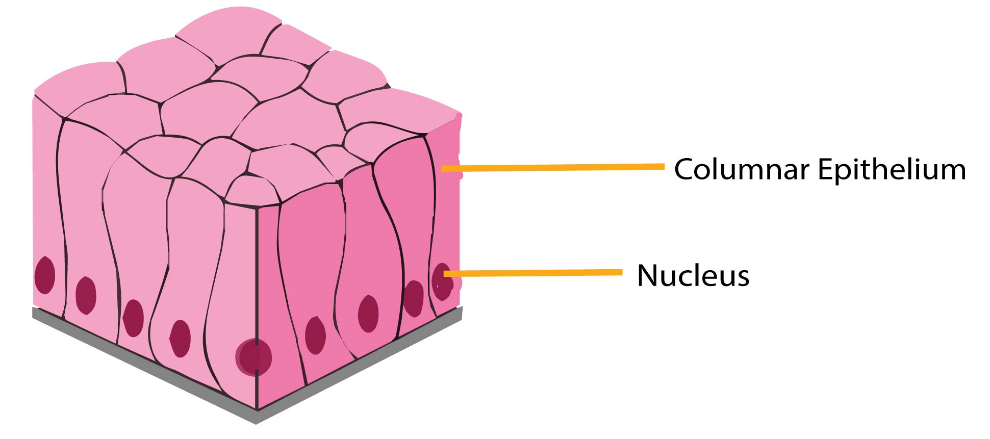
Simple Epithelial Tissue Is Organised As A Single Layer Of Cells And Stratified Epithelial Tissue Is Formed By Several Layers Of Cells.
Related Post:
