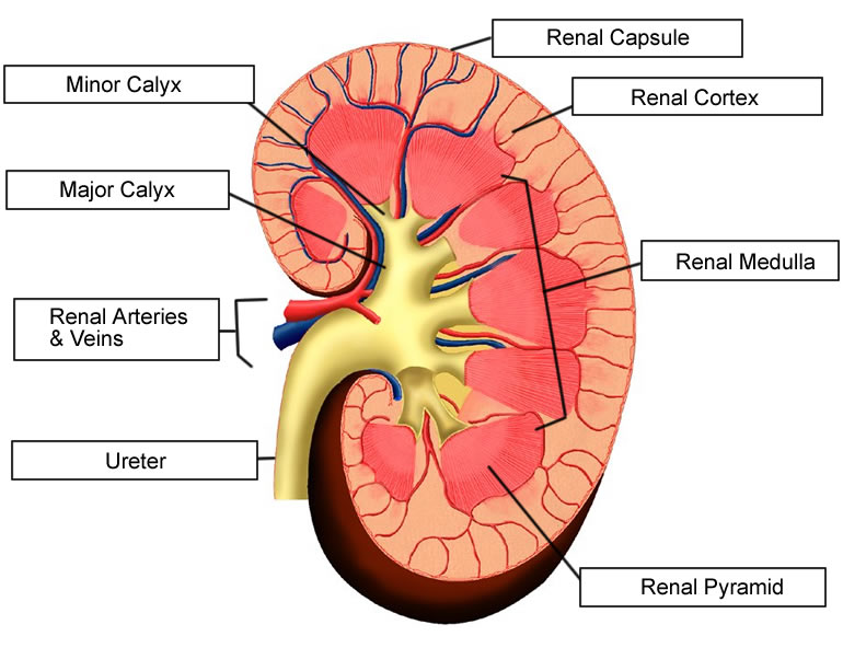
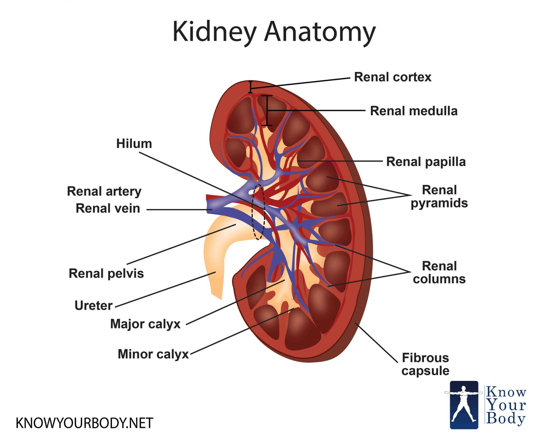
Label The Schematic Drawing Of A Kidney
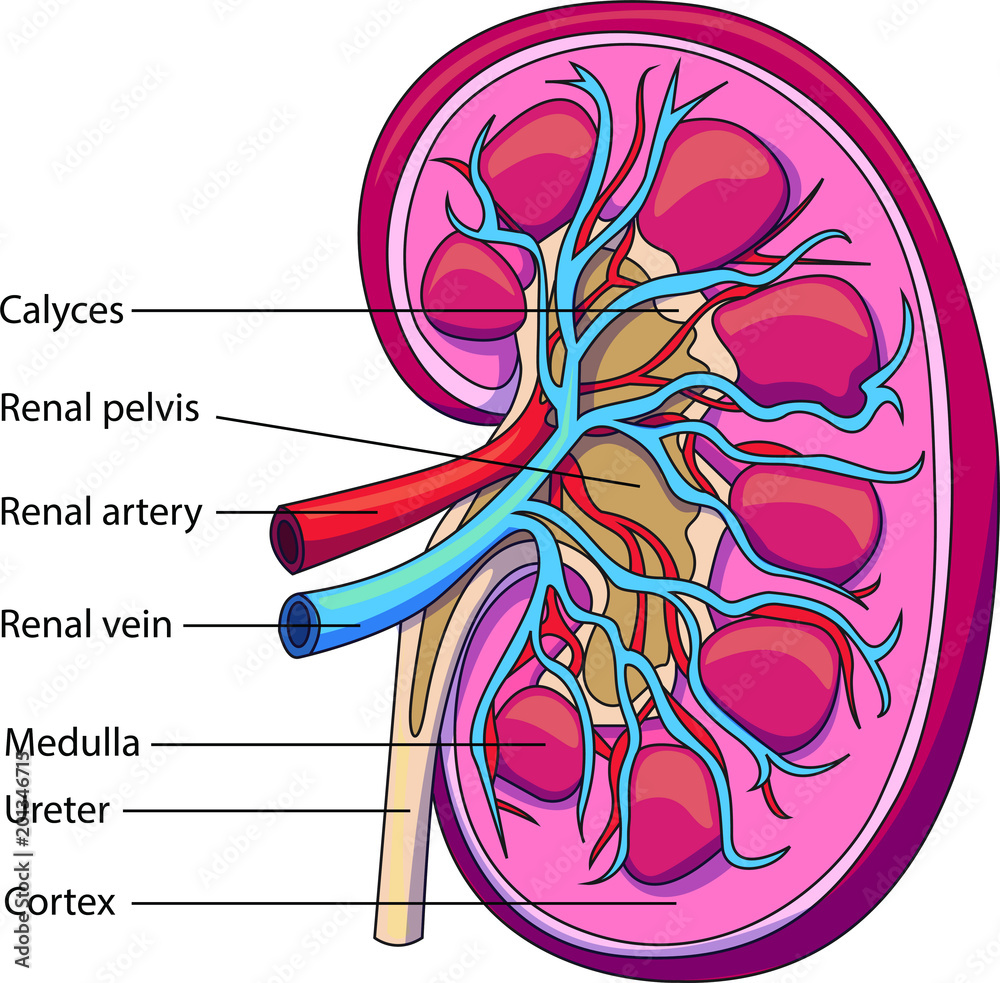
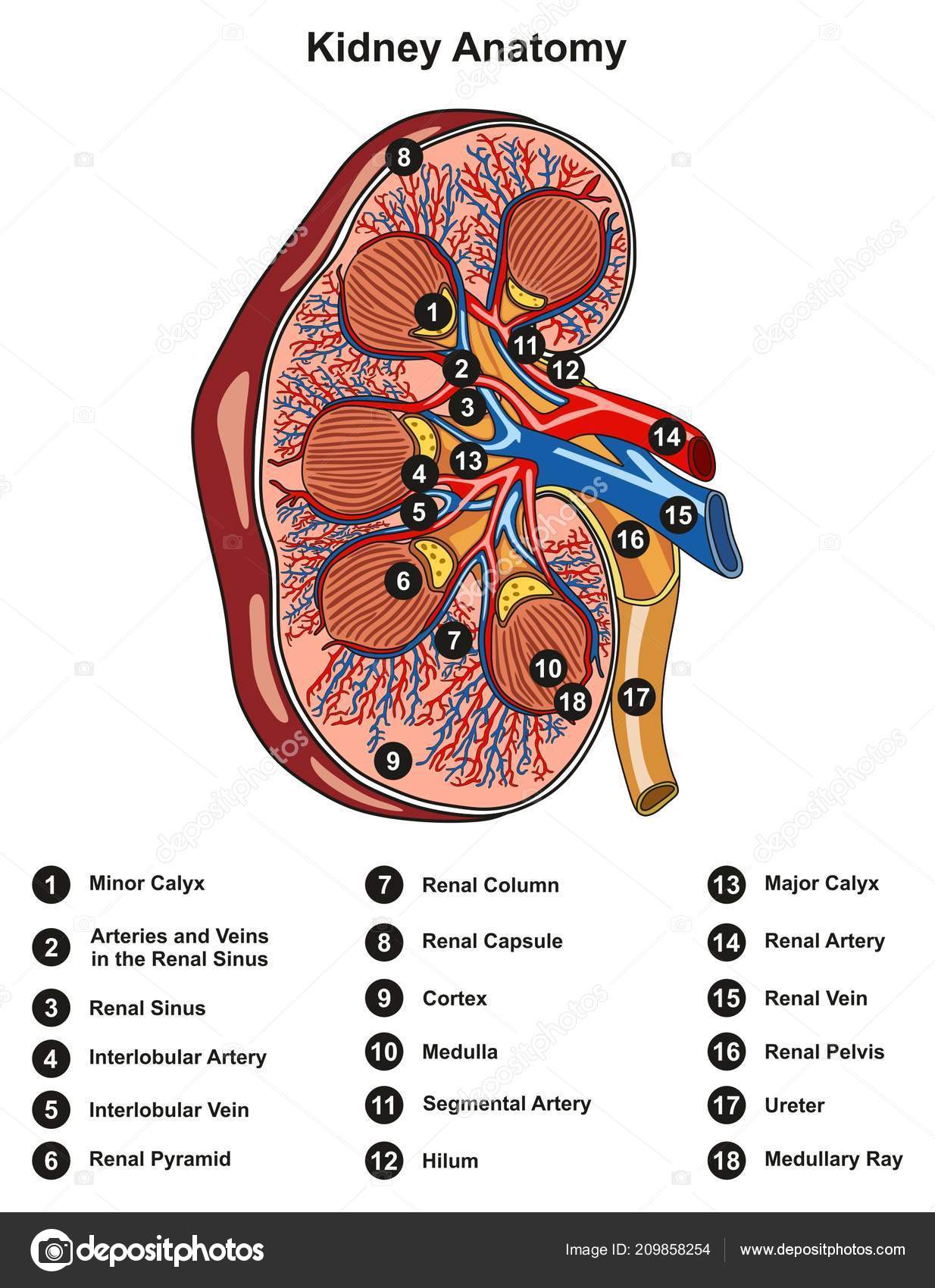
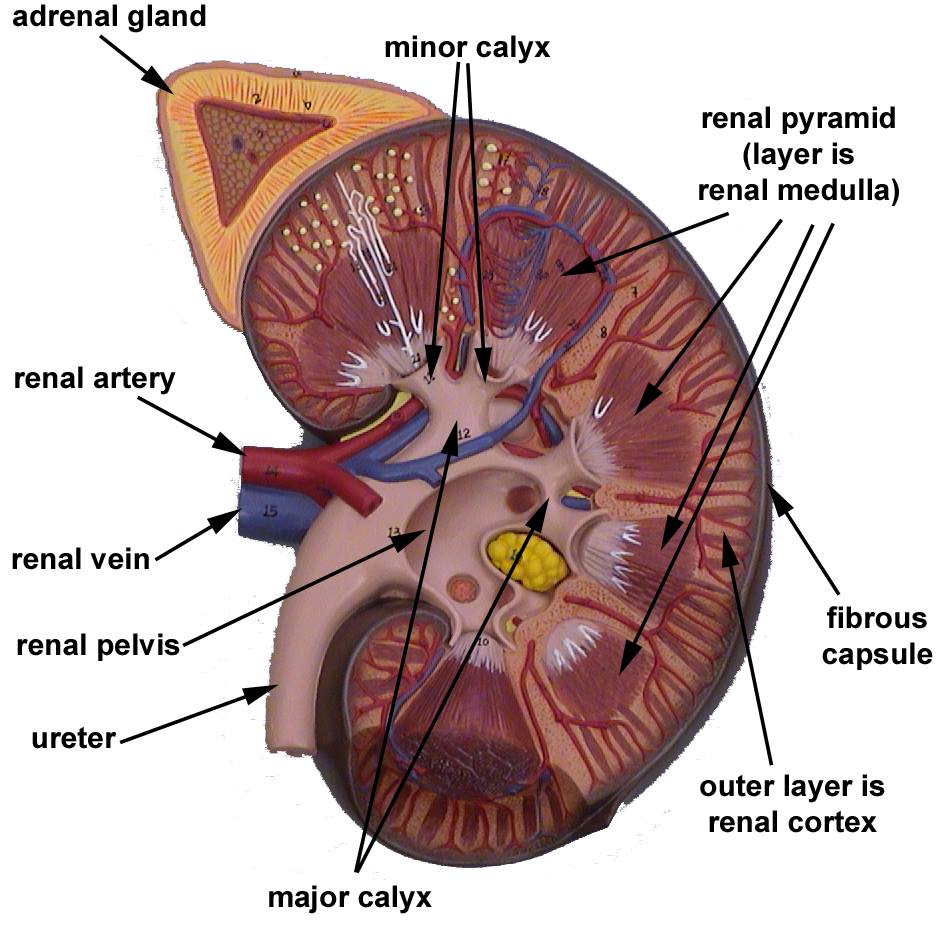
Label The Schematic Drawing Of A Kidney - Identify the major internal divisions and structures of the kidney; Web health tips what are kidneys? (2 ½ inches) wide and 3 cm. Each kidney looks like the kidney bean and the renal hilum is the entry and exit site for structures servicing the kidneys: Web label the kidney by mpurzycki +1 27,965 plays 9 questions ~30 sec english 9p more 9 too few (you: Remaining 0 correct 0 wrong 0 press play! Web the question provides us with a diagram that represents a human kidney. Web nephron, functional unit of the kidney, the structure that actually produces urine in the process of removing waste and excess substances from the blood. Urethra minor calyx renal pelvis renal medulla renal pyramid ureter renal cortex major calyx this problem has been solved! In males and 135 gms in females. Web each kidney consists of a cortex, medulla and calyces. Learn vocabulary, terms, and more with flashcards, games, and other study tools. A small curvature called renal hilum is present on the concave face of the kidney from where the renal artery enters the kidney, and renal vein and ureter leave the kidney. Web this article covers the anatomy of. The waste fluid then travels through a series of tubules where water and electrolytes will be reabsorbed into the body. Try the fastest way to create flashcards Also, the diagram shows the relationship between the. In males and 135 gms in females. Web label the kidney by mpurzycki +1 27,965 plays 9 questions ~30 sec english 9p more 9 too. Try the fastest way to create flashcards Learn vocabulary, terms, and more with flashcards, games, and other study tools. A small curvature called renal hilum is present on the concave face of the kidney from where the renal artery enters the kidney, and renal vein and ureter leave the kidney. The waste fluid then travels through a series of tubules. Try the fastest way to create flashcards Web the first slide is an overview of the urinary system that shows the kidneys, ureters, urinary bladder, and urethra. Web label the kidney by mpurzycki +1 27,965 plays 9 questions ~30 sec english 9p more 9 too few (you: Each kidney looks like the kidney bean and the renal hilum is the. Web label the kidney by mpurzycki +1 27,965 plays 9 questions ~30 sec english 9p more 9 too few (you: Urethra minor calyx renal pelvis renal medulla renal pyramid ureter renal cortex major calyx this problem has been solved! Web on the superior aspect of each kidney is an adrenal gland. They also help filter blood before sending it back. Web health tips what are kidneys? Each kidney looks like the kidney bean and the renal hilum is the entry and exit site for structures servicing the kidneys: Web label the kidney by mpurzycki +1 27,965 plays 9 questions ~30 sec english 9p more 9 too few (you: Web label the schematic drawing of a kidney. Inside this capsule, two. The average weight of adult kidney is about 150 gms. Web label the schematic drawing of a kidney. Web the kidneys are the body's filter system. Web microscopic structure in the cortex of the kidney; Web this diagram shows where the renal artery enters the kidney, and where the renal vein leaves. Surrounds the glomerulus collects the waste fluid filtered out of the glomerulus. Students can practice labeling the structures and color coding the diagram. Inside this capsule, two distinct regions can be observed: Web label the schematic drawing of a kidney. Web kidneys are dark brown in colour and are embedded in a mass of fat. Surrounds the glomerulus collects the waste fluid filtered out of the glomerulus. Before starting, ensure that you have all the necessary materials to label the kidney diagram. Web this article covers the anatomy of the kidneys, their function and internal structure together with the nephron. They produce urine to carry the wastes out of the body. In this article we. Learn more and see the diagrams at kenhub! Also, the diagram shows the relationship between the. The kidney is packed with around a million structures called nephrons close nephron filtration unit of. Remaining 0 correct 0 wrong 0 press play! Web microscopic structure in the cortex of the kidney; The kidney is packed with around a million structures called nephrons close nephron filtration unit of. Urethra minor calyx renal pelvis renal medulla renal pyramid ureter renal cortex major calyx this problem has been solved! Surrounds the glomerulus collects the waste fluid filtered out of the glomerulus. Students drag labels to the structures on the slide. Web start studying correctly label the following anatomical parts of a kidney. Web kidneys are dark brown in colour and are embedded in a mass of fat. (1 ½ inch) in thickness. Students can practice labeling the structures and color coding the diagram. (2 ½ inches) wide and 3 cm. Compare and contrast the cortical and juxtamedullary nephrons Web it clearly shows the locations of the right and left kidney, as well as the large blood vessels that connect the kidneys to the body’s main artery (aorta) and vein (inferior vena cava). Learn vocabulary, terms, and more with flashcards, games, and other study tools. In this article we will explore the microanatomy of a nephron and learn how their function relates to their histological features. Web the question provides us with a diagram that represents a human kidney. Web microscopic structure in the cortex of the kidney; Each kidney looks like the kidney bean and the renal hilum is the entry and exit site for structures servicing the kidneys:
Kidney Structures Learn Surgery Online

Draw the L.S of kidney and label the parts.

Human kidney medical diagram with a cross section Vector Image

Please send me a diagram of L.S Of kidney and label the main parts of
labelled diagram of human kidney
Schematic vector diagram of a kidney. Kidney structure with labeled
Labeled Kidney Diagram World of Reference
Biology (MBBS) Structure of Human Kidney with labeled diagram Ratta.pk
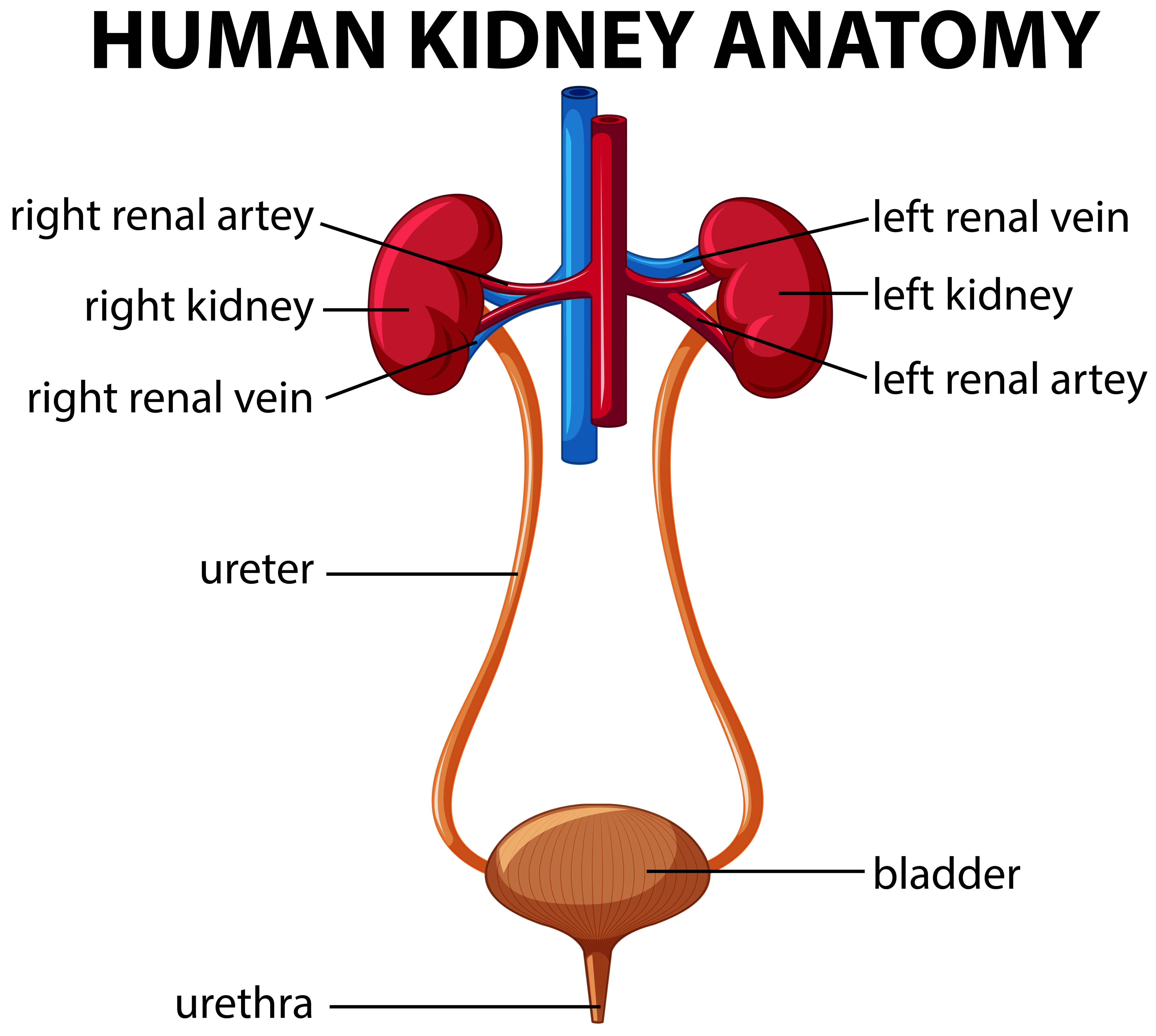
Label the Parts of the Urinary System
Human Body Organs Diagram Kidneys Human Body Anatomy
0% 08:00.0 Other Games Of Interest
Learn Vocabulary, Terms, And More With Flashcards, Games, And Other Study Tools.
Web Start Studying Kidney Anatomy Labeling.
Remaining 0 Correct 0 Wrong 0 Press Play!
Related Post: