Muscle Cell Drawing
Muscle Cell Drawing - Web a muscle cell, also known as a myocyte, is a mature contractile cell in the muscle of an animal. These cells can be found in various forms, including skeletal, cardiac, and smooth muscle tissues, each serving distinct functions within the body. So, get productive and begin drawing these cells. Web how to draw muscle cell step by step. You will find some unique features in cardiac muscle that will help you to differentiate it from. Skeletal muscle cells compose the muscle cells linked to manhood and therefore are significant in locomotion. Cardiac muscle cells are branched and striated, but short. All three muscle tissues have some properties in common; Skeletal, smooth, and cardiac (cardiomyocytes). Web the cell bodies of some pns neurons, such as the motor neurons that control skeletal muscle (the type of muscle found in your arm or leg), are located in the cns. [2] a skeletal muscle cell is long and threadlike with many nuclei and is called a muscle fiber. Compare and contrast the functions of each muscle tissue type. Skeletal muscle fibers can be quite large compared to other cells, with diameters up to 100 μ m and lengths up to. Figure 8.2 components of a skeletal muscle. Most organisms are. Web figure 10.4 muscle fiber a skeletal muscle fiber is surrounded by a plasma membrane called the sarcolemma, which contains sarcoplasm, the cytoplasm of muscle cells. These motor neurons have long extensions (axons) that run from the cns all the way to the muscles they connect with (innervate). Web these cell drawing ideas will make you learn how to label. They all exhibit a quality called excitability as their plasma membranes can change their electrical states. Muscle cell diagram, how to draw smooth muscle cell, how to draw. Most popular hand drawn seamless pattern vector seamless. Compare and contrast the functions of each muscle tissue type. Skeletal muscle fibers can be quite large compared to other cells, with diameters up. Web 16/10/2023 17/12/2022 by anatomylearner the cardiac muscle under a microscope shows a short cylindrical fiber with a centrally placed oval nucleus. One can draw cells from muscle memory after going over these cell drawing ideas. Muscle cell diagram, how to draw smooth muscle cell, how to draw. Most organisms are multicellular and have cells that are specialised to do. Web a muscle cell, also known as a myocyte, is a mature contractile cell in the muscle of an animal. Muscle cells, commonly called myocytes, would be the cells that cosmetics muscle tissue. Web a muscle cell, known technically as a myocyte, is a specialized animal cell which can shorten its length using a series of motor proteins specially arranged. Web muscle is one of the four primary tissue types of the body, and the body contains three types of muscle tissue: These cells can be found in various forms, including skeletal, cardiac, and smooth muscle tissues, each serving distinct functions within the body. This is due to their extensive length and appearance. Cardiac and skeletal myocytes are occasionally known. What is your request drawing? While several associated proteins help, actin and myosin form thick and thin filaments which slide past each other to contract small units of a muscle cell. Skeletal, smooth, and cardiac (cardiomyocytes). They all exhibit a quality called excitability as their plasma membranes can change their electrical states. Skeletal muscle, smooth muscle, and cardiac muscle, visualized. Muscle cell diagram, how to draw smooth muscle cell, how to draw. Other materials in each muscle include nerve cells, collagen and elastin fibers, fat, and blood vessels ( figure 8.2 ). Smooth muscle cells are short, tapered at each end, and have only one plump nucleus in each. Skeletal, smooth, and cardiac (cardiomyocytes). Skeletal muscle, cardiac muscle, and smooth. A muscle fiber is composed of many fibrils, which give the cell its striated appearance. Web a muscle cell, or myocyte, is a specialized animal cell designed for contraction, facilitated by organized motor proteins, primarily actin and myosin. Web how to draw muscle cell step by step. These motor neurons have long extensions (axons) that run from the cns all. Muscle cells are commonly called myocytes. A muscle cell is a long cell as compared to other kinds of cells, and many muscle cells connect with each other to create the long fibers present in muscle tissue. Figure 8.2 components of a skeletal muscle. Web because skeletal muscle cells are long and cylindrical, they are commonly referred to as muscle. Smooth muscle cells are short, tapered at each end, and have only one plump nucleus in each. Web a muscle cell, also known as a myocyte, is a mature contractile cell in the muscle of an animal. Muscle cell diagram, how to draw smooth muscle cell, how to draw. Most popular hand drawn seamless pattern vector seamless. A muscle cell is a long cell as compared to other kinds of cells, and many muscle cells connect with each other to create the long fibers present in muscle tissue. Skeletal muscle, smooth muscle, and cardiac muscle, visualized here using light microscopy. Skeletal muscle fibers can be quite large compared to other cells, with diameters up to 100 μ m and lengths up to. Cardiac and skeletal myocytes are occasionally known as muscle fibers because of their lengthy and fibrous form. Muscle cells are commonly called myocytes. What is your request drawing? Skeletal muscle, cardiac muscle, and smooth muscle ( figure 10.2 ). They are contractile, meaning they can shorten and generate a pulling force. Skeletal muscle cells compose the muscle cells linked to manhood and therefore are significant in locomotion. Web figure 10.4 muscle fiber a skeletal muscle fiber is surrounded by a plasma membrane called the sarcolemma, which contains sarcoplasm, the cytoplasm of muscle cells. So let me actually draw it really a lot bigger here. These contain thick and thin myofilaments made up mainly of the proteins actin and myosin.
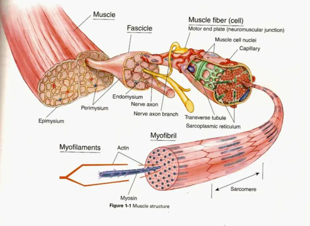
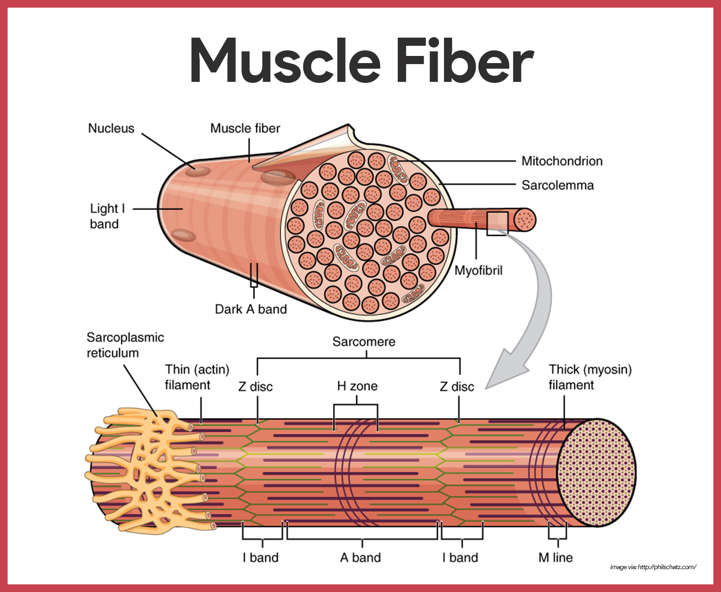
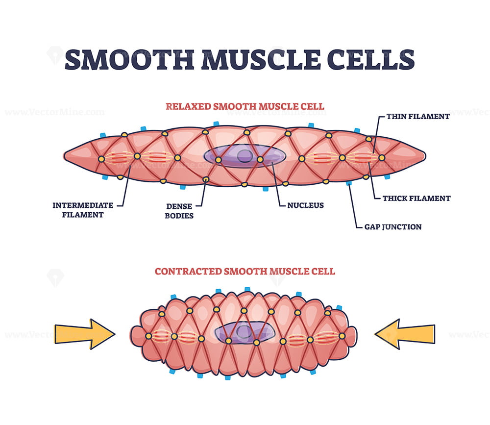
Muscle Skeletal Muscle Cell A hand drawn sketch by Dr. Ch… Flickr

Anatomy/Muscular System Science Olympiad Student Center Wiki
Muscle cell diagram

Types of muscle cell diagram 1762350 Vector Art at Vecteezy
Muscular System Anatomy and Physiology Nurseslabs
Diagram Showing Types Of Muscle Cells Stock Illustration Download

Schematic representation of the skeletal muscle structure. The

Anatomy Of Muscle Cell The Anatomy Stories
Smooth Muscle Cell Structure

Types of muscle cells vector illustration Types of muscles, Tissue
Cardiac Muscle Cells Are Branched And Striated, But Short.
A Muscle Fiber Is Composed Of Many Fibrils, Which Give The Cell Its Striated Appearance.
Compare And Contrast The Functions Of Each Muscle Tissue Type.
Web 16/10/2023 17/12/2022 By Anatomylearner The Cardiac Muscle Under A Microscope Shows A Short Cylindrical Fiber With A Centrally Placed Oval Nucleus.
Related Post:
