Elastic Connective Tissue Drawing
Elastic Connective Tissue Drawing - But elastic fibers are present in relatively high concentration in several organs, including the largest arteries in the body. This image shows a portion of the wall of the aorta, the large vessel that carries. Web elastic fibers provide flexibility to the tissues. Connective tissue can be classified as connective tissue proper, cartilage, bone, or blood. This fiber consists of thin strands of collagen that form a network of fibers to support the. Web this image of loose connective tissue shows collagenous fibers (red), elastic fibers (black), matrix, and fibroblasts (cells that produce the fibers). In loose connective tissue, the fibers are loosely organized, leaving large spaces in between. Blood and haemopoietic tissue 7. This tissue is most widely distributed connective tissue in the animal body. Fill out the blanks next to your drawing. Web dense connective tissue is reinforced by bundles of fibers that provide tensile strength, elasticity, and protection. Like all tissue types, it consists of cells surrounded by a compartment of fluid called the extracellular matrix (ecm). The fibers and other components of the connective tissue matrix are secreted by fibroblasts. Web identify and distinguish between the types of connective tissue:. Like all tissue types, it consists of cells surrounded by a compartment of fluid called the extracellular matrix (ecm). Connective tissue can be classified as connective tissue proper, cartilage, bone, or blood. Dense regular connective tissue fibers are parallel to each other, enhancing tensile strength and resistance to stretching in the direction of the fiber orientations. Loose connective tissue, also. Supportive connective tissue —bone and cartilage—provide structure and strength to the body and protect soft tissues. As may be obvious from its name, one of the major functions of connective tissue is to connect tissues and organs. As its name implies, connective tissue is a term given to several body tissues that connect, support, and help bind other tissues. It. Ed reschke/photolibrary/getty images in vertebrates, the most common type of connective tissue is loose connective tissue. The image summarizes the various categories of connective tissues found in the human body. Web elastic connective tissue 40x human aorta c.s. Loose connective tissue, also called areolar connective tissue, has a sampling of all of the components of a connective tissue. Blood and. This is especially seen in the arterial blood vessels and walls of the bronchial tubes. Web in drawing images of connective tissue proper preparations seen under the microscope, it is important to simplify the visuals. Elastic fibers differ from collagen in. Web the following points highlight the ten main varieties of connective tissues of human body. Ed reschke/photolibrary/getty images in. Web elastic fibers provide flexibility to the tissues. But elastic fibers are present in relatively high concentration in several organs, including the largest arteries in the body. Blood and haemopoietic tissue 7. Web identify and distinguish between the types of connective tissue: Web figure 10.3.5 10.3. Explain the functions of connective tissues. Ed reschke/photolibrary/getty images in vertebrates, the most common type of connective tissue is loose connective tissue. The fibers and other components of the connective tissue matrix are secreted by fibroblasts. Supportive connective tissue —bone and cartilage—provide structure and strength to the body and protect soft tissues. (a) areolar tissue (= loose connective tissue): Web there are connective tissue fibroblast and blood vessels in the outer layer of the perichondrium of elastic cartilage histology. Web introduction connective tissue is one of the basic tissue types of the body. Osseous tissue or bone 10. Ligaments and tendons are mostly formed from dense regular connective tissue. Web structure and cellular components of loose connective tissue. Web elastic connective tissue. The image summarizes the various categories of connective tissues found in the human body. This fiber consists of thin strands of collagen that form a network of fibers to support the. Connective tissue can be classified as connective tissue proper, cartilage, bone, or blood. Supportive connective tissue —bone and cartilage—provide structure and strength to the body. Other components include collagen fibers (c) and elastic fibers (ef) • observe the characteristics of the three types of muscle The main fibers that form this tissue are elastic in nature. Explain the functions of connective tissues. Web obtain a slide of ground compact bone connective tissue from the slide box. Explain the functions of connective tissues. Web structure and cellular components of loose connective tissue. Web elastic connective tissue 40x human aorta c.s. Web the following points highlight the ten main varieties of connective tissues of human body. Loose connective tissue with nuclei (n) labeled. • observe the characteristics of the three types of muscle It is an important structural element that supports and separates spaces between our organs and tissues in the human body. As its name implies, connective tissue is a term given to several body tissues that connect, support, and help bind other tissues. Reticular fibers are the third type of protein fiber found in connective tissues. In the circle below, draw a representative sample of key features you identified, taking care to correctly and clearly draw their true shapes and directions. Web elastic fibers provide flexibility to the tissues. Elastic fibers differ from collagen in. But elastic fibers are present in relatively high concentration in several organs, including the largest arteries in the body. True elastic connective tissue is very rare, and we have no slide specimens that show it. These fibers allow the tissues to recoil after stretching. It comprises a diverse group of cells that can be found in different parts of the body.Elastic Connective Tissue Stock Photo Download Image Now iStock

dense elastic connective tissue Nurse Midwife Anatomy, physiology
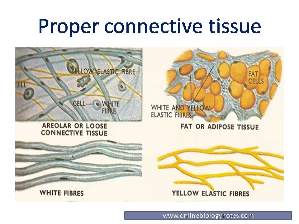
Proper connective tissue Areolar, Adipose, Reticular, white fibrous
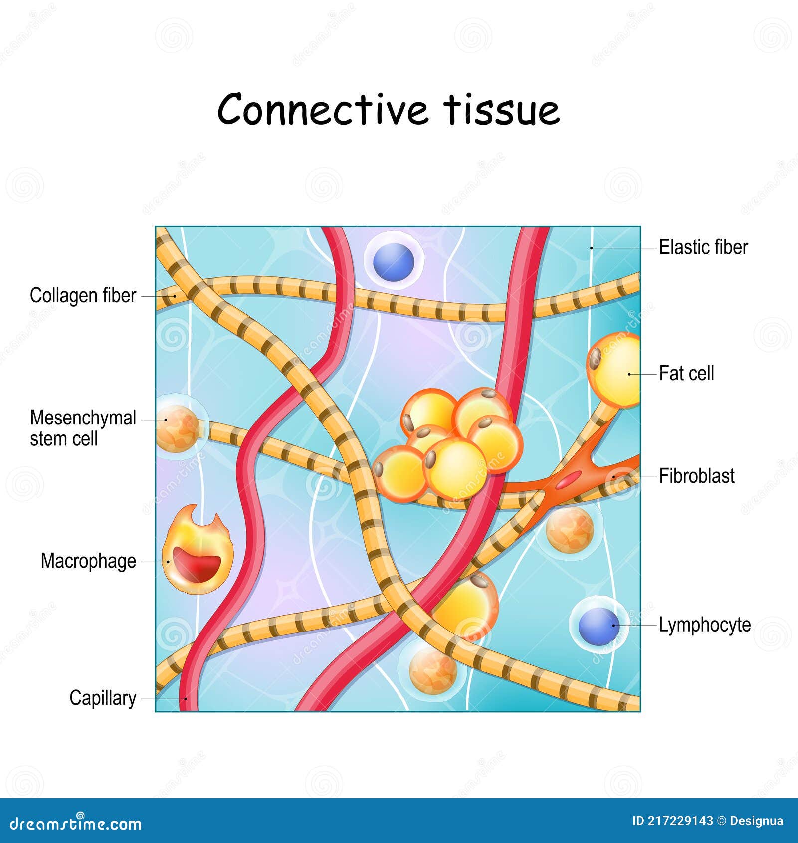
Connective Tissue. Structure and Anatomy Stock Vector Illustration of
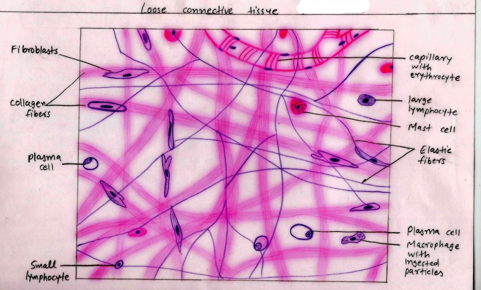
Histology Image Connective tissue

PPT Connective Tissues PowerPoint Presentation, free download ID

A&P1 Lab 3 elastic connective tissue YouTube
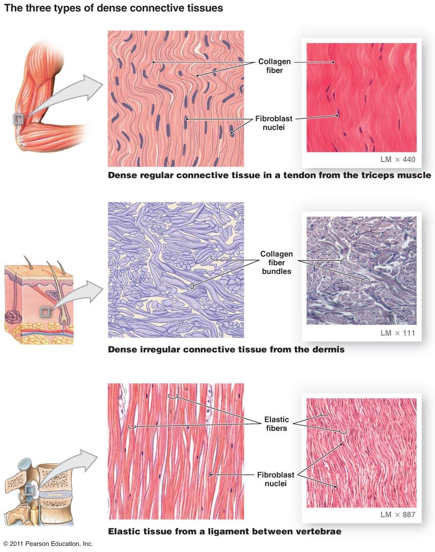
Connective Tissue; Structure and Function McIsaac Health Systems Inc.

Dense Regular Elastic Connective Diagram Quizlet
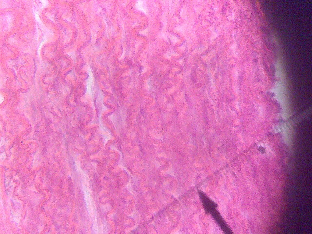
Elastic connective tissue Science, anatomy ShowMe
• Study The Characteristics Of Loose, Dense, Elastic, And Reticular Connective Tissue, Adipose Tissue, Cartilage, And Bone.
Other Components Include Collagen Fibers (C) And Elastic Fibers (Ef)
Ed Reschke/Photolibrary/Getty Images In Vertebrates, The Most Common Type Of Connective Tissue Is Loose Connective Tissue.
The Image Summarizes The Various Categories Of Connective Tissues Found In The Human Body.
Related Post:
