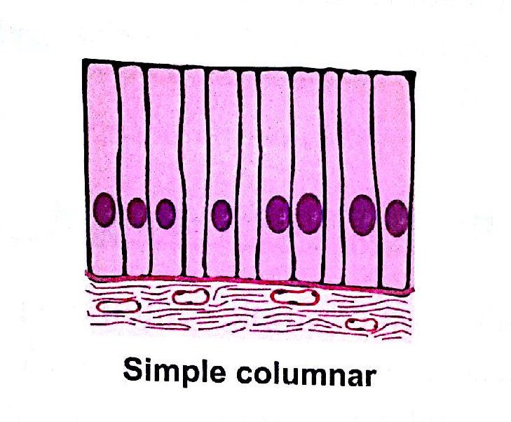
Simple Columnar Drawing
Simple Columnar Drawing - These cells, shaped like cubes, have microvilli and cilia that help move the fluid around. Again, if you want to see these simple columnar epithelium as the verticle or transverse section,. Ependymal cells, glial cells of the central nervous system, form a barrier between cerebral spinal fluid and interstitial fluid. A cuboidal epithelial cell looks close to a square. Instead of being smooth, the inside of the intestine is folded and covered by millions of tiny projections called villi. Web there are three basic shapes used to classify epithelial cells. Allows absorbtion, secretes mucous and enzymes pseudostratified columnar location: Web distinguish between simple epithelia and stratified epithelia, as well as between squamous, cuboidal, and columnar epithelia describe the structure and function of endocrine and exocrine glands and their respective secretions A columnar epithelial cell looks like a column or a tall rectangle. Web simple columnar epithelial cells can specialize to secret mucus that coats and protects the surrounding surface from damage (insert link). Web diya's drawing channel 805 subscribers subscribe 2.1k views 1 year ago #how_to_draw_epithelial_tissue welcome to diya's art tutorial youtube channel. Web the simple columnar epithelium is a type of epithelium that is formed of a single layer of long, elongated cells mostly in areas where absorption and secretion are the main functions. Web simple columnar location: Like cuboidal epithelium, the. Bodytomy provides a labeled diagram to help you understand the structure and function of simple columnar epithelium. Again, if you want to see these simple columnar epithelium as the verticle or transverse section,. The cells of this epithelium are arranged in a neat row. These cells, shaped like cubes, have microvilli and cilia that help move the fluid around. Simple. This type of epithelium is adapted for secretion and/or absorption, and can also be protective. Allows absorbtion, secretes mucous and enzymes pseudostratified columnar location: Bodytomy provides a labeled diagram to help you understand the structure and function of simple columnar epithelium. • how to draw simple columnar epitheliu. Simple secretory columnar epithelium lines the stomach and uterine cervix.the simple columnar. Because the epithelium can be innervated, simple columnar epithelium is also specialized to provide sensory input. Web simple columnar epithelium: Web primate small intestine you are looking at part of the small intestine. Web distinguish between simple epithelia and stratified epithelia, as well as between squamous, cuboidal, and columnar epithelia describe the structure and function of endocrine and exocrine glands. Bodytomy provides a labeled diagram to help you understand the structure and function of simple columnar epithelium. Web there are three basic shapes used to classify epithelial cells. Web simple epithelium is one of the types of epithelium that is divided into simple columnar epithelium, simple squamous epithelium, and simple cuboidal epithelium. Web diya's drawing channel 805 subscribers subscribe 2.1k. This type of epithelium is adapted for secretion and/or absorption, and can also be protective. Simple secretory columnar epithelium lines the stomach and uterine cervix.the simple columnar epithelium that lines the intestine also contains a few goblet cells. Bodytomy provides a labeled diagram to help you understand the structure and function of simple columnar epithelium. Web simple columnar epithelia are. Instead of being smooth, the inside of the intestine is folded and covered by millions of tiny projections called villi. Web #biology #simplecolunar #ciliatedcolumnar #columnarepithelium #class9 #class11 #hscbiology #maharashtrastateboard2021 #biology2021 #biologydiagrams #icse #cb. The cells of this epithelium are arranged in a neat row. Web distinguish between simple epithelia and stratified epithelia, as well as between squamous, cuboidal, and columnar. The cells of this epithelium are arranged in a neat row. Web simple columnar epithelia are tissues made of a single layer of long epithelial cells that are often seen in regions where absorption and secretion are important features. This type of epithelium is adapted for secretion and/or absorption, and can also be protective. These cells, shaped like cubes, have. Like cuboidal epithelium, the cells in the columnar epithelium are also modified to suit the function and structure of the organ better. Web about transcript this video describes the structure and function of ependymal cells. Web simple columnar epithelia are tissues made of a single layer of long epithelial cells that are often seen in regions where absorption and secretion. Simple secretory columnar epithelium lines the stomach and uterine cervix.the simple columnar epithelium that lines the intestine also contains a few goblet cells. Read more about modified simple columnar epithelium, dog jejenum, 20x; Web there are three basic shapes used to classify epithelial cells. Again, if you want to see these simple columnar epithelium as the verticle or transverse section,.. The inner surface of the intestinal wall is made of simple columnar epithelium (sce). Read more about modified simple columnar epithelium, dog jejenum, 20x; Web #biology #simplecolunar #ciliatedcolumnar #columnarepithelium #class9 #class11 #hscbiology #maharashtrastateboard2021 #biology2021 #biologydiagrams #icse #cb. Web the stomach wall, with simple columnar epithelium visible as a lining at the top. Trachea and most of the upper respiratory tract (ciliated cells) Web the simple columnar epithelium is a type of epithelium that is formed of a single layer of long, elongated cells mostly in areas where absorption and secretion are the main functions. Like the cuboidal epithelia, this epithelium is active in the absorption and secretion of molecules using active transport. This type of epithelium is adapted for secretion and/or absorption, and can also be protective. A squamous epithelial cell looks flat under a microscope. Web diya's drawing channel 805 subscribers subscribe 2.1k views 1 year ago #how_to_draw_epithelial_tissue welcome to diya's art tutorial youtube channel. Web primate small intestine you are looking at part of the small intestine. Web simple columnar location: Web simple columnar epithelium: Ependymal cells, glial cells of the central nervous system, form a barrier between cerebral spinal fluid and interstitial fluid. The cells of this epithelium are arranged in a neat row. Web tall columnar epithelium lines the ducts of many exocrine glands.
How to draw Pseudostratified columnar epithelium most easy way
simple columnar Medical Study Zone
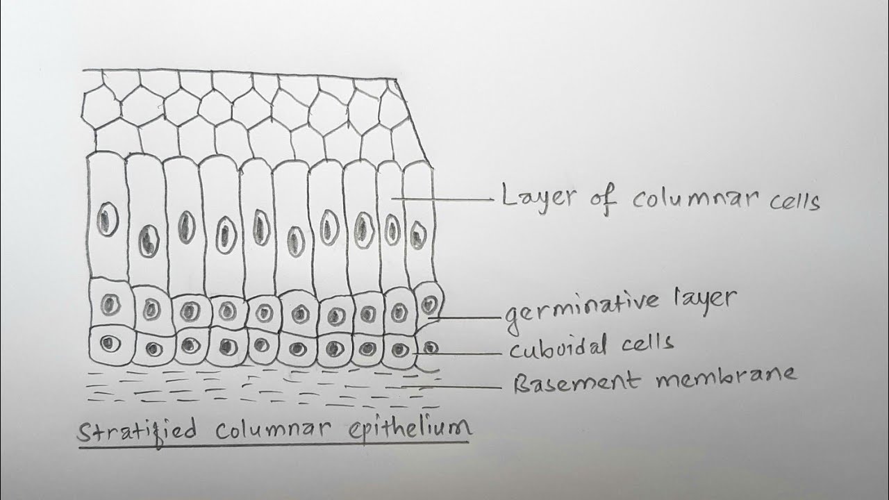
How to draw stratified columnar epithelium easy way YouTube
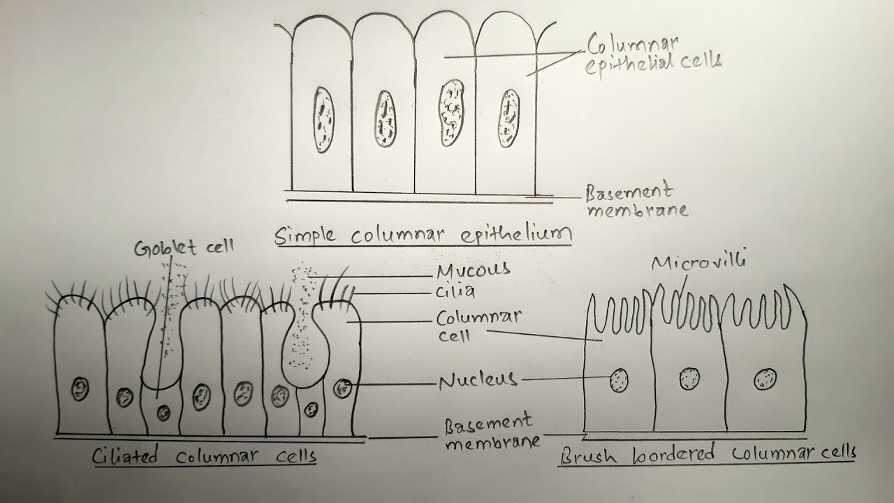
How to draw simple columnar epithelium types of comlumnar epithelium

Simple Columnar Tutorial Histology Atlas for Anatomy and Physiology
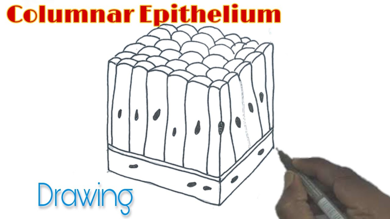
Columnar Epithelium Drawing/ How to draw Columnar Epithelium YouTube

Simple Columnar Tutorial Histology Atlas for Anatomy and Physiology

Drawing of Simple Columnar Tissue Calligraphy, Drawings, Tissue

Simple columnar Simple columnar epithelium, Tissue types, Types of

Simple Columnar Tutorial Histology Atlas for Anatomy and Physiology
Bodytomy Provides A Labeled Diagram To Help You Understand The Structure And Function Of Simple Columnar Epithelium.
These Cells, Shaped Like Cubes, Have Microvilli And Cilia That Help Move The Fluid Around.
• How To Draw Simple Columnar Epitheliu.
A Columnar Epithelial Cell Looks Like A Column Or A Tall Rectangle.
Related Post: